A molecular life in four acts
There’s a point in the life of a skin cell when preserving one of its proteins would prevent it from receiving something it needs. The protein has already helped reorganise the cell’s interior, but its next contribution depends on being dismantled. Its amino acids, and the compounds made from them, will help the cell hold water after it reaches the surface.
The protein is filaggrin. It begins as a much larger precursor, passes through a brief existence as an intact molecule, then supplies some of the small, water-retaining compounds found in the outer epidermis. Each change makes a different kind of behaviour possible. A molecule that interacts with cellular fibres cannot do the same job as the soluble products released from its breakdown.
In this way, I think of filaggrin as a sacrificial protein. That’s my way of describing its biology, rather than a formal class of protein: part of its contribution to the barrier depends on surrendering the molecular form that performed the job before it. The word also needs a little restraint. This is controlled biochemistry, and the location and timing of destruction are as consequential as the destruction itself.
Following this sequence takes us from a living cell to the material it leaves behind at the surface, and eventually into the chemistry of skin acidity. Before we get to the acids, though, the skin has to assemble an unusually large protein.
ACT I: The reserve
The surface of your skin is built from cells that have completed a substantial conversion. Deeper in the epidermis, keratinocytes are living cells, with nuclei, organelles and the machinery to synthesise new proteins. As they migrate toward the surface they undergo differentiation, where they prepare to lose much of that machinery. Their destination is the stratum corneum, where flattened cells called corneocytes are embedded in an extracellular lipid barrier.
A corneocyte has lost the equipment that would let it respond like a living keratinocyte. It can no longer metabolise or produce new proteins, and any further processes are carried out by enzymes it has inherited from the keratinocyte, but it still has a physical job. It must withstand the harsh conditions at the surface without becoming excessively brittle or allowing the barrier to fall apart. Some of the chemistry the corneocyte will later require is prepared in advance while its predecessor, the keratinocyte, can still make proteins.
In the granular layer, near the top of the living epidermis, keratinocytes produce profilaggrin. The prefix ‘pro’ marks a precursor: a large protein containing repeated sections that will later be released as individual filaggrin units. The human FLG gene commonly encodes around 10 to 12 of these repeats, plus terminal regions with their own properties. A single precursor therefore contains the material for several future molecules, connected within the same chain.[1]
Partners of the head. Bunick’s group found three proteins binding the profilaggrin N-terminus: annexin II, stratifin and HSP27. Annexin II needed calcium to bind; stratifin did not, and it sat alongside profilaggrin in the granular layer of human skin.[2]
The precursor also has a calcium-binding region at its N-terminus, the end of the chain where the protein begins. Christopher Bunick and colleagues resolved this S100 domain as a pair of matching subunits, each binding two calcium ions.[2] This gives us a close view of one folded part of the precursor, separate from the repeated sections that will become mature filaggrin. In the appendix model it is the region marked in gold, a folded head on a chain that is otherwise largely flexible. Calcium also changes what this head does. In solution at high calcium, the domain opened up to expose hydrophobic patches and clumped into larger assemblies, and Bunick’s group proposed that calcium helps pack profilaggrin into the storage granules described below.[2]
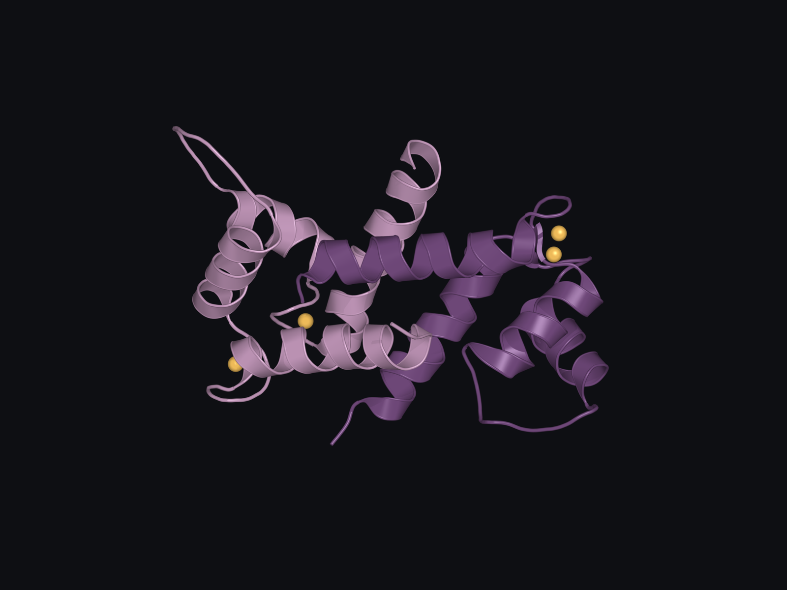
Within living granular cells, the repeat region is heavily phosphorylated. Phosphate groups are attached to amino-acid side chains, altering the protein’s charge while its future units remain connected.[2] Extensive phosphorylation of the repeats, alongside calcium binding at the head, has been proposed as a requirement for packing the precursor into granules.[2] The phosphates also add negative charge to units whose later grip on keratin is thought to depend on positive charge, a contrast Acts II and III return to.
Profilaggrin accumulates in structures called keratohyalin granules. Seen in an image of the epidermis, these look satisfyingly solid: conspicuous bodies inside a cell, apparently holding a supply until it’s needed. Their physical behaviour makes that description incomplete.
Human keratinocytes and mouse epidermis. The experiments combined engineered proteins in cultured human keratinocytes with observations in mouse epidermis. The pH-sensitive behaviour concerns conditions inside the cell.[3]
In 2020, work from Elaine Fuchs’s laboratory showed that filaggrin-containing granules behave as liquid-like condensates. Experiments with repeat-containing proteins in human keratinocytes, supported by observations in mouse epidermis, identified liquid-liquid phase separation as the driver of their assembly.[3] A concentrated protein-rich phase forms within the surrounding cellular fluid, without needing a membrane to enclose it. The familiar comparison is droplets separating from a mixture, although a living cell imposes considerably more control over its droplets than a bottle of salad dressing does.
The repeats contribute to that behaviour. In the experimental system, a single filaggrin unit did not form the condensates produced by longer repeat-containing constructs; terminal regions also altered their properties. The precursor’s organisation therefore affects what the protein does before those units are released.
These granules also occupy a crowded cellular environment. The researchers found that keratin bundles constrained their ability to merge, connecting the behaviour of the condensates to the surrounding cytoskeleton. They also linked pH-sensitive granule dynamics to terminal differentiation. This concerns conditions inside the cell; it shouldn’t be confused with the acidity measured at the skin’s surface.
The precursor phase already has a story of its own. Profilaggrin is concentrated within an organised structure whose behaviour depends on the protein’s sequence and its surroundings. As the keratinocyte approaches cornification, that arrangement changes, and the repeated sections become available in a different molecular form.
ACT II: A change of form
Cornification converts a living keratinocyte into a corneocyte. The nucleus and organelles disappear, an armoured protein envelope develops, and lipids are supplied to the space outside the cell. The resulting cell has to function as barrier material after it has lost the capacity to maintain itself in its usual way.
Profilaggrin processing is part of that conversion. The precursor is stripped of its phosphate groups and cleaved by proteases, enzymes that cut peptide bonds within a protein chain. Individual mature filaggrin units are then released from the larger molecule. Cleavage here produces a protein with a new opportunity to act.
Mature filaggrin goes on to interact with intermediate filaments, including keratin, the protein fibres that give epidermal cells much of their internal mechanical organisation. Filaggrin’s name itself comes from “filament-aggregating protein”. It describes a capacity that becomes especially apparent when researchers introduce the mature protein into cells experimentally.
Beverly Dale, Richard Presland and their colleagues did that in the 1990s, expressing human filaggrin in cultured cells. Intermediate-filament networks collapsed into aggregates, nuclear integrity was disrupted and the cells rounded up and lost attachment to the substratum. Precursor-like constructs containing repeated units, meanwhile, formed granules.[4] Different forms of the same protein family produced very different cellular behaviour.
Those were artificial expression experiments, so they don’t establish that filaggrin alone flattens a normal human keratinocyte. What they do show is that the mature protein has a considerable capacity to reorganise filamentous networks. In cornification, that capacity becomes part of a coordinated programme in which the cell’s internal organisation is already being transformed.
That helps explain why the seemingly inactive precursor deserves attention. Preparing a supply of connected repeats is biologically different from releasing the mature protein into the cytoplasm, and the epidermis keeps the two states apart. Granule organisation and precursor processing govern the transition between them. But why go to the trouble? The evidence points toward an answer. A protein that can collapse a keratin network would have very different consequences in a living cell that still depends on its keratin network to hold its shape, and holding its release back until cornification is under way would be consistent with that.
The protease SASPase offers one experimental route into this control. Mice lacking it accumulated abnormal profilaggrin-processing products, had less mature filaggrin and developed a thicker, less hydrated stratum corneum.[5] Interfering with release changed the tissue that eventually formed at the surface.
There’s a revealing complication in this experiment: the reported free-amino-acid composition was unchanged. A processing defect therefore cannot automatically be assumed to cause a shortage of downstream moisturising compounds. Filaggrin has consequences at more than one stage, and different interruptions do not produce identical outcomes.
An unexpected ancestry. SASPase arose from a retroviral sequence that the genome domesticated long ago. It still resembles HIV-1 protease closely enough that one HIV drug, indinavir, binds it, although its hydrophobic pockets make it resistant to most of the others.[6]
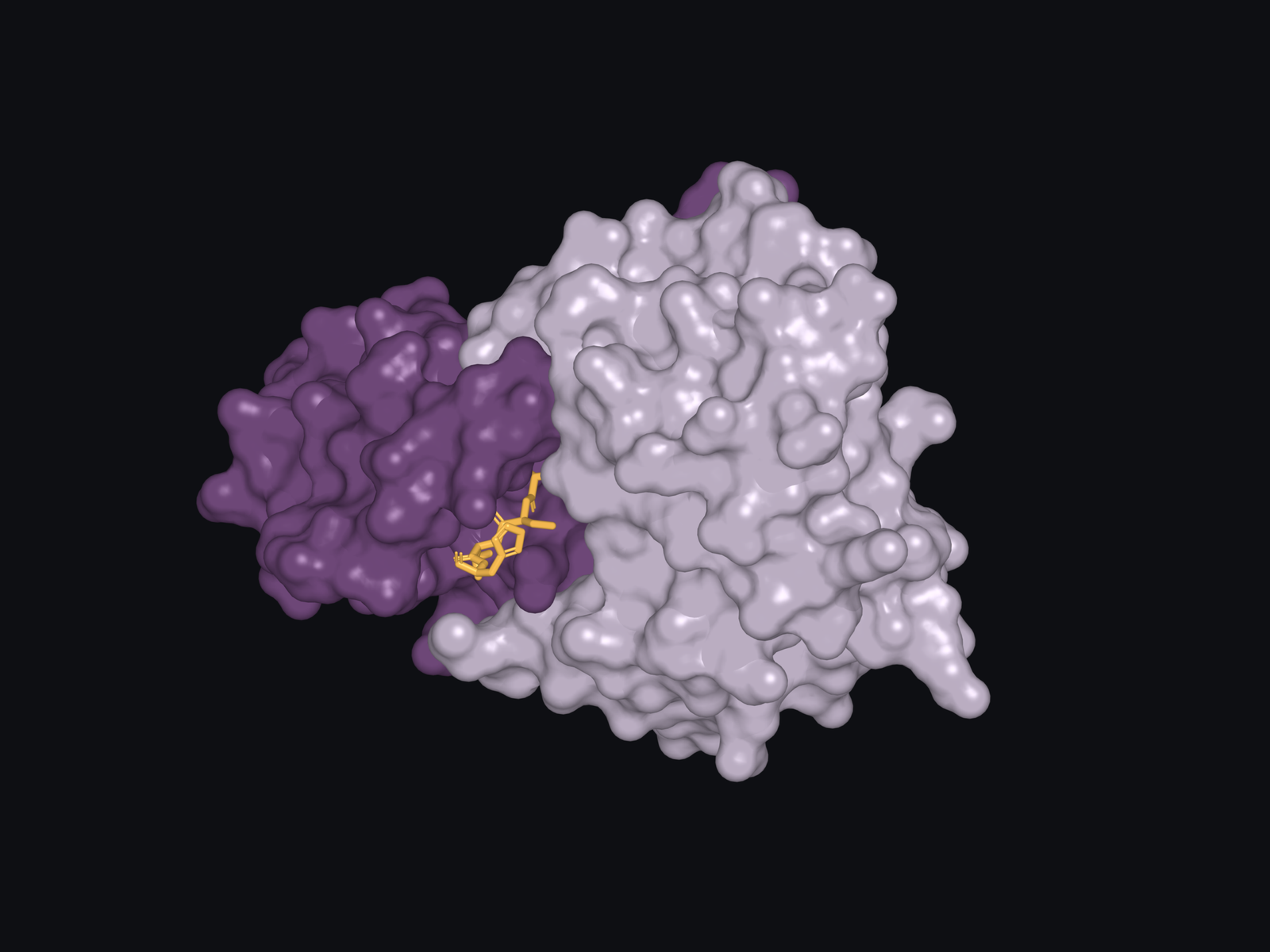
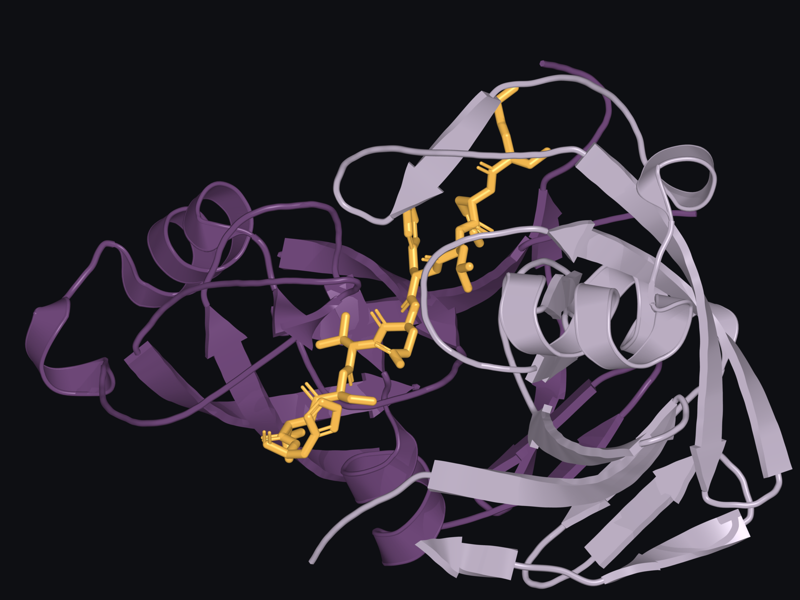
Structural work brings us closer to the recognition behind processing. Profilaggrin is a chain of near-identical repeats, so releasing intact units requires careful cutting of the linkers between them, not through the units themselves. Matsui’s group showed that recombinant SASPase cuts a linker peptide directly.[5] SASPase, also called ASPRV1, forms a pair of subunits around a cleft that accommodates a short filaggrin-derived peptide. Xueqian Feng and colleagues showed how hydrophobic pockets within SASPase, which accommodate water-avoiding side chains, strictly require hydrophobic residues on either side of the cut, which narrows where along the chain the enzyme can act.[6] A protease’s access to a chain is only part of the story; the sequence it encounters also shapes the outcome.
The structure below captures binding using a catalytically inactive enzyme. It shows how a small section of the substrate can fit within the protease, giving us a molecular view of recognition during the release stage.[6]
For the molecules that proceed towards breakdown, the next transition presents another chemical problem. Filaggrin’s interactions with keratin have been valuable during reorganisation, but much of it is destined to become free-floating, soluble material within the corneocyte. Before proteases can finish that conversion, the protein’s charge takes another turn.
ACT III: Letting go
Filaggrin contains many arginine residues. An amino acid becomes a residue when it is incorporated into a protein chain, and arginine’s side chain carries a positive charge under physiological conditions. Together, the abundance of arginine residues contributes to filaggrin’s strongly basic character and its interactions with keratin. In the ionic zipper model proposed by Mack, Steven and Steinert, its positively charged regions attract negatively charged sites on keratin filaments, helping filaggrin bind to and draw the fibres together.[7]
Enzymes called peptidylarginine deiminases, or PADs, convert protein-bound arginine into citrulline. Unlike arginine’s side chain, citrulline’s side chain is neutral. The reaction, called deimination or citrullination, alters the charge carried by the protein without cutting its backbone, leading to changes in its chemical interactions. This is the second charge change in filaggrin’s life. Dephosphorylation stripped negative charge as the units were released; deimination now removes positive charge as they let go.
PAD1 and PAD3 have been implicated in epidermal filaggrin deimination through studies of their location and their activity on protein substrates.[8] This gives us a candidate mechanism for modifying filaggrin before extensive degradation. In this model, loss of positive charge weakens the chemical basis of its association with keratin and changes the substrate that the breakdown enzymes encounter.
It’s easy to compress everything after this point into ‘filaggrin is degraded’, but that phrase oversimplifies a cascade of multiple reactions, all operating at different scales. A protease can cut a large protein into smaller peptides, each still containing several amino acids. Freeing individual amino acids from those fragments requires further processing. Stripping the positive charge from an arginine side chain is a different job again, done by a different enzyme.
Biochemical experiments by Yayoi Kamata and colleagues, using bleomycin hydrolase purified from newborn rat epidermis, separated some of those steps. Their work examined deiminated filaggrin and implicated calpain-1 in generating peptide fragments, with bleomycin hydrolase releasing amino acids from peptide substrates. Caspase-14 produced a different cleavage pattern.[9] The experiments make the sequence of cuts more tangible, although results with isolated substrates don’t establish one compulsory order for every enzyme in human skin.
A different molecular fate. Cross-linking joins proteins through covalent bonds within the corneocyte’s tough envelope. Filaggrin material incorporated there becomes part of that structure. Its presence in the envelope does not tell us what proportion follows this route.[10]
We can follow productive dismantling without knowing the complete sequence. Deimination changes the protein’s interactions; proteolysis breaks its chain; further processing makes small compounds available. Some filaggrin material is incorporated into the cross-linked cornified envelope.[10] The sequence we’re following here concerns the material processed into free amino acids; but it doesn’t describe the final destination of every molecule.
The strongest test of the sacrificial idea comes from interrupting the later stages while allowing filaggrin to form.
Mice lacking caspase-14. This experiment tests what happens when later filaggrin processing is interrupted.[11]
In 2011, Esther Hoste and colleagues studied mice lacking caspase-14. Their epidermis still processed profilaggrin into filaggrin repeat units. The initial release had occurred, but filaggrin fragments accumulated, and two important downstream products, pyrrolidone carboxylic acid and urocanic acid, were substantially reduced.[11]
The result separated production from completion. Having filaggrin present doesn’t guarantee that the corneocyte will contain its later chemical products. The chain has to be successfully processed for those products to become available. In this mouse model, interrupting degradation left more intermediate material and less of what should have followed.
This is why the destruction is a part of filaggrin’s functional life. The caspase-14 experiment makes the idea more than an attractive reading of the pathway: a failure to complete breakdown deprives the tissue of measurable outputs. Once we ask what happens to those outputs, the protein’s apparent disappearance becomes easier to understand, and harder to describe as a loss.
ACT IV: What remains
Named for what it does. Natural moisturising factor is named for its job, not its source. Filaggrin supplies much of the amino-acid fraction; lactate, urea and salts reach the mixture by other routes.
The stratum corneum contains a mixture of small, water-retaining compounds called natural moisturising factor, usually shortened to NMF. Much of this material is held inside corneocytes, where it contributes to their ability to hold water. The mixture includes free amino acids and derivatives, as well as components such as lactate, urea and salts. Filaggrin breakdown supplies an important part of it.
These small soluble molecules behave differently from their parent protein that associated with keratin. Their interaction with water contributes to hydration within the cell, allowing the stratum corneum to retain flexibility as it meets the outside environment. A corneocyte cannot synthesise a new protein in response to a dry afternoon. Its ability to cope therefore depends on material prepared and processed during its formation.
Two filaggrin-derived products deserve their full chemical names, because their routes into this mixture vary.
Pyrrolidone carboxylic acid, or PCA, forms through cyclisation of glutamine released during filaggrin breakdown. This reaction can occur without an enzyme: the amino group closes onto the side chain to form a ring, and ammonia is released. PCA is strongly water-attracting, so this conversion turns part of filaggrin’s amino-acid supply into a form primed for retaining water. Early experiments in guinea-pig epidermis traced its formation to protein-derived material and supported nonenzymatic cyclisation of free glutamine as the predominant route.[12]
Histidine takes another route. The enzyme histidase, also called histidine ammonia-lyase, converts free histidine into trans-urocanic acid, or UCA.[13] This reaction also releases ammonia from an amino acid that has been freed from the protein.
Neither reaction acts on the intact protein. Both work on amino acids already freed from the chain, which separates them from the earlier PAD reaction, where arginine was modified while it was still part of the chain. The breakdown has now supplied precursors for two acids, both of which contribute to the chemical environment of the stratum corneum.
The supply is also responsive to conditions around the epidermis. In 2017, Laura Cau’s group grew reconstructed human epidermis at different relative humidities, comparing a very humid condition above 95% with a drier condition of 30–50%.[14] The tissue developed under drier conditions had more NMF, together with changes in stratum corneum structure and filaggrin processing.
One result explains why the earlier charge-changing reaction was worth following. PAD1 expression and protein deimination increased in the drier tissue, without an increase in the measured expression or activity of the breakdown proteases. A PAD inhibitor partly reversed the processing response. The inhibitor wasn’t selective for PAD1, but the authors suggested that deimination plays a major role in how humidity drives filaggrin breakdown.
The system had altered the substrate presented to its enzymes. An increase in processing didn’t require a corresponding increase in every enzyme that could cut the protein. This was epidermis developed under controlled humidity conditions, so it doesn’t give us a perfect timetable for what happens to someone’s skin after they walk into a dry room. What it does show is that the conversion from protein to small compounds responds to the environment in which the barrier forms. The epidermis was adapting to its environment.
That brings us to acidity. Filaggrin supplies precursors for acids, including UCA, within the stratum corneum. It is tempting to draw a direct line from less filaggrin to fewer acids to a higher surface pH. The first step is well supported in relevant experiments. The final step depends on the skin’s acidifying system taken as a whole.
Skin has overlapping routes for establishing acidity, and the rest of this series will show several of them. With NHE1 and lipid-derived acids feeding the same environment, removing one source was unlikely to neutralise the surface. In experiments using mice with defective histidase activity or filaggrin processing, loss of the expected filaggrin-histidine-UCA contribution did not produce the straightforward loss of acidity predicted by an obligatory pathway.[15] The result tells us the route isn’t essential. It doesn’t tell us it’s unimportant, and the chemical contribution that filaggrin supplies still remains.
Reconstructed human skin. Filaggrin was reduced in a laboratory-grown skin construct. The compensatory acidifying responses were measured under those controlled conditions.[16]
A reconstructed-skin experiment from Kateřina Vávrová’s laboratory makes the consequences of compensation especially clear. Reducing filaggrin lowered PCA and UCA, yet surface pH remained acidic. Expression of NHE1, the sodium-hydrogen exchanger, increased. By day 14, secretory phospholipase A₂-IIA activity had also risen and free fatty acids had doubled, providing another route into acidification.[16]
The acidity held. It came, however, with less ordered lipid layers and increased permeability, which the authors linked to the excess fatty acid. The tissue had kept a familiar output while changing how it arrived there. A surface pH reading therefore couldn’t tell the whole story of what had happened underneath.
This is where filaggrin’s contribution connects to the broader acidity series. NHE1 exchanges sodium and hydrogen ions (protons) across a membrane; lipid-processing enzymes provide a different source of acidic products. Those mechanisms will have their own future explanations. Filaggrin reaches this shared chemical environment through the transformation of a protein, with hydration and cell structure already part of its history.
When that history is interrupted at the beginning, the consequences extend beyond losing one mature molecule. Loss-of-function variants in FLG cause ichthyosis vulgaris, a condition characterised by dry, scaly skin.[17] Reduced precursor production limits the material available to enter the sequence, affecting the opportunity for both intact filaggrin and its later products to act.
Inflammation provides another route to a reduced supply. In experiments using keratinocytes, the type 2 cytokines IL-4 and IL-13 suppressed filaggrin expression, together with observations of reduced expression in atopic dermatitis.[18] A person can therefore have less filaggrin available without the explanation being confined to an inherited change in its gene.
Human measurements connect these routes to the products at the surface. Sanja Kezic’s team studied 96 Irish children with moderate-to-severe atopic dermatitis and 24 controls, measuring filaggrin breakdown products in tape-stripped skin.[19] Levels were influenced by FLG genotype and disease severity. Children with atopic dermatitis who lacked the screened mutations also had lower levels than controls.
In this study, the NMF measurement was defined as PCA plus UCA, rather than the entire mixture. Even with that narrower measure, the result gives the molecular story a human consequence: the supply of downstream compounds varies with both inherited filaggrin status and the disease environment. The cultured-cell cytokine experiments provide one mechanism through which inflammation can reduce the supply, although the human measurements alone can’t assign every reduction to those cytokines.
By the time a corneocyte reaches the surface, much of the filaggrin that passed through it has ceased to exist as filaggrin. Its afterlife remains in the cell’s small-molecule chemistry, including compounds that retain water and feed into an overlapping system of acidification.
That is the sense in which the protein gives itself away.
Appendix Understanding the low confidence model
This larger render below offers a view of how the precursor is represented by an AlphaFold model. It covers residues 1–1400 of the 4061-residue profilaggrin reference sequence, so it represents part of profilaggrin. The loops and turns are the model’s proposed coordinates. They are not an experimentally observed arrangement of the precursor inside a granule.[20]
AlphaFold reports a local confidence score called pLDDT for each residue, on a scale from 0 to 100. The mean for this model is 38.27, and 90.7% of its residues score 50 or below. That is low confidence across most of the model. The score describes confidence in the predicted local structure; it is not a percentage probability that the whole molecule is either correct or incorrect.[20,21]
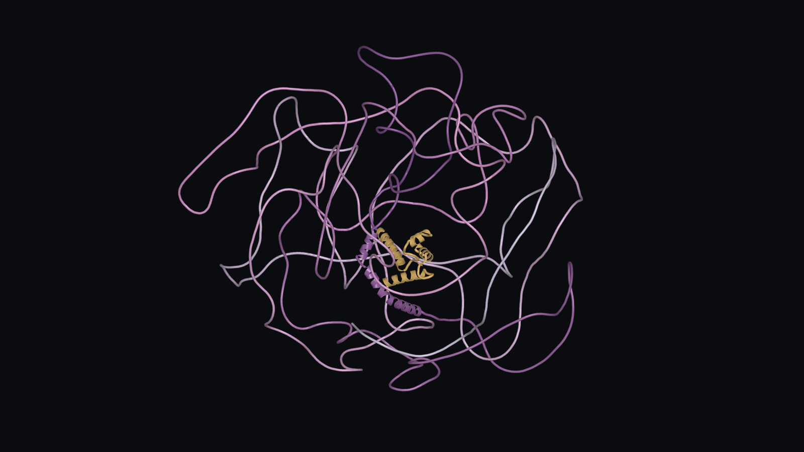
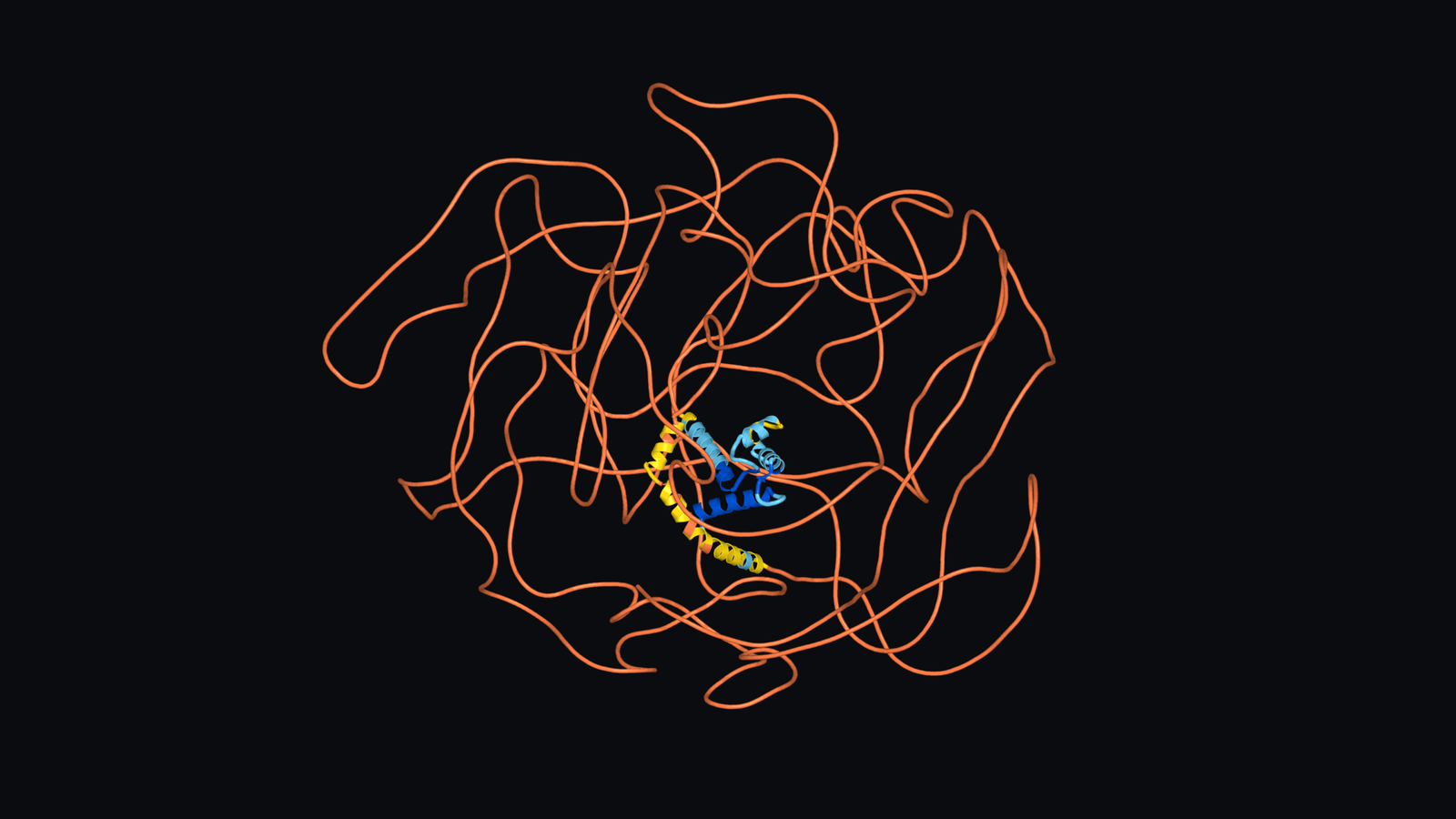
Low pLDDT can arise from flexibility or intrinsic disorder, or from insufficient information for a reliable prediction.[21] That in itself can be informative, as intrinsically disordered regions (IDRs) are important in their own right. An intrinsically disordered region explores a range of conformations instead of maintaining one stable fold, often participating in dynamic biological interactions. The score alone cannot distinguish those explanations or demonstrate phase separation. We can experimentally resolve profilaggrin’s folded S100 domain, while its surrounding repeat regions require further evidence about their changing conformations.
The connection to condensates comes from experiments by Quiroz and colleagues, where they examined filaggrin’s disordered repeat-containing sequences and demonstrated how repeat number and terminal regions affected condensate formation.[3] The results make the flexibility biologically relevant. A protein can contribute to an organised cellular structure through interactions between many chains, even when those chains do not each maintain one fixed shape.
An individual flexible chain can change conformation. A condensate’s collective interactions allow many molecules to assemble, exchange material and change their physical behaviour. The granule experiments connected that behaviour to keratin organisation and intracellular pH as cells differentiated.[3] Resolving these flexible regions therefore requires evidence about a range of conformations and the conditions that alter them. The render supplies one uncertain arrangement, helping us discuss a dynamic protein while keeping its experimentally observed behaviour in sight.[3,20,21]
Profilaggrin. The large precursor protein containing connected filaggrin repeats, which are later released through processing.
Keratohyalin granules. Protein-rich structures in granular-layer keratinocytes where profilaggrin accumulates before cornification.
Condensates. Concentrated assemblies of molecules that form a distinct phase within the surrounding cellular fluid without an enclosing membrane. The granules discussed here show liquid-like behaviour.
Cornification. The specialised conversion of an epidermal keratinocyte into a corneocyte, including loss of its nucleus and organelles and formation of a tough protein envelope.
Deimination. Conversion of protein-bound arginine into citrulline. This changes the side chain’s charge without cutting the protein backbone; it is also called citrullination.
Proteolysis. Cleavage of peptide bonds by enzymes called proteases, breaking a protein or peptide into smaller pieces.
Cornified envelope. The tough, covalently cross-linked protein structure formed around the interior of a corneocyte during cornification.
Natural moisturising factor. A mixture of small, water-retaining compounds within corneocytes. Filaggrin-derived amino acids and their products contribute to the mixture, alongside substances from other sources.
References
1. Gan, S.Q., McBride, O.W., Idler, W.W., et al. (1990). Organization, structure, and polymorphisms of the human profilaggrin gene. Biochemistry, 29(40), pp. 9432–9440. https://doi.org/10.1021/bi00492a018
2. Bunick, C.G., Presland, R.B., Lawrence, O.T., Pearton, D.J., Milstone, L.M., and Steitz, T.A. (2015). Crystal Structure of Human Profilaggrin S100 Domain and Identification of Target Proteins Annexin II, Stratifin, and Hsp27. Journal of Investigative Dermatology, 135(7), pp. 1801–1809. https://doi.org/10.1038/jid.2015.102
3. Quiroz, F.G., Fiore, V.F., Levorse, J., et al. (2020). Liquid-liquid phase separation drives skin barrier formation. Science, 367(6483), eaax9554. https://doi.org/10.1126/science.aax9554
4. Dale, B.A., Presland, R.B., Lewis, S.P., et al. (1997). Transient expression of epidermal filaggrin in cultured cells causes collapse of intermediate filament networks with alteration of cell shape and nuclear integrity. Journal of Investigative Dermatology, 108(2), pp. 179–187. https://doi.org/10.1111/1523-1747.ep12334205
5. Matsui, T., Miyamoto, K., Kubo, A., et al. (2011). SASPase regulates stratum corneum hydration through profilaggrin-to-filaggrin processing. EMBO Molecular Medicine, 3(6), pp. 320–333. https://doi.org/10.1002/emmm.201100140
6. Feng, X., Chen, Z., Lan, C., and Ding, J. (2026). Structure and enzymatic properties of human retroviral-like aspartic protease 1 and functional roles of disease-associated mutations. Acta Biochimica et Biophysica Sinica. Published online 11 August 2026. https://doi.org/10.3724/abbs.2026141. Structure record: https://www.rcsb.org/structure/27IQ
7. Mack, J.W., Steven, A.C. and Steinert, P.M. (1993). The mechanism of interaction of filaggrin with intermediate filaments. The ionic zipper hypothesis. Journal of Molecular Biology, 232(1), pp. 50–66. https://doi.org/10.1006/jmbi.1993.1369
8. Méchin, M.C., Enji, M., Nachat, R., et al. (2005). The peptidylarginine deiminases expressed in human epidermis differ in their substrate specificities and subcellular locations. Cellular and Molecular Life Sciences, 62(17), pp. 1984–1995. https://doi.org/10.1007/s00018-005-5196-y
9. Kamata, Y., Taniguchi, A., Yamamoto, M., et al. (2009). Neutral cysteine protease bleomycin hydrolase is essential for the breakdown of deiminated filaggrin into amino acids. Journal of Biological Chemistry, 284(19), pp. 12829–12836. https://doi.org/10.1074/jbc.M807908200
10. Steinert, P.M. and Marekov, L.N. (1995). The proteins elafin, filaggrin, keratin intermediate filaments, loricrin, and small proline-rich proteins 1 and 2 are isodipeptide cross-linked components of the human epidermal cornified cell envelope. Journal of Biological Chemistry, 270(30), pp. 17702–17711. https://doi.org/10.1074/jbc.270.30.17702
11. Hoste, E., Kemperman, P., Devos, M., et al. (2011). Caspase-14 is required for filaggrin degradation to natural moisturizing factors in the skin. Journal of Investigative Dermatology, 131(11), pp. 2233–2241. https://doi.org/10.1038/jid.2011.153
12. Barrett, J.G. and Scott, I.R. (1983). Pyrrolidone carboxylic acid synthesis in guinea pig epidermis. Journal of Investigative Dermatology, 81(2), pp. 122–124. https://doi.org/10.1111/1523-1747.ep12542975
13. Scott, I.R. (1981). Factors controlling the expressed activity of histidine ammonia-lyase in the epidermis and the resulting accumulation of urocanic acid. Biochemical Journal, 194(3), pp. 829–838. https://doi.org/10.1042/bj1940829
14. Cau, L., Pendaries, V., Lhuillier, E., et al. (2017). Lowering relative humidity level increases epidermal protein deimination and drives human filaggrin breakdown. Journal of Dermatological Science, 86(2), pp. 106–113. https://doi.org/10.1016/j.jdermsci.2017.02.280
15. Fluhr, J.W., Elias, P.M., Man, M.-Q., et al. (2010). Is the filaggrin-histidine-urocanic acid pathway essential for stratum corneum acidification? Journal of Investigative Dermatology, 130(8), pp. 2141–2144. https://doi.org/10.1038/jid.2010.74
16. Vávrová, K., Henkes, D., Strüver, K., et al. (2014). Filaggrin deficiency leads to impaired lipid profile and altered acidification pathways in a 3D skin construct. Journal of Investigative Dermatology, 134(3), pp. 746–753. https://doi.org/10.1038/jid.2013.402
17. Smith, F.J.D., Irvine, A.D., Terron-Kwiatkowski, A., et al. (2006). Loss-of-function mutations in the gene encoding filaggrin cause ichthyosis vulgaris. Nature Genetics, 38(3), pp. 337–342. https://doi.org/10.1038/ng1743
18. Howell, M.D., Kim, B.E., Gao, P., et al. (2007). Cytokine modulation of atopic dermatitis filaggrin skin expression. Journal of Allergy and Clinical Immunology, 120(1), pp. 150–155. https://doi.org/10.1016/j.jaci.2007.04.031
19. Kezic, S., O’Regan, G.M., Yau, N., et al. (2011). Levels of filaggrin degradation products are influenced by both filaggrin genotype and atopic dermatitis severity. Allergy, 66(7), pp. 934–940. https://doi.org/10.1111/j.1398-9995.2010.02540.x
20. RCSB Protein Data Bank (n.d.). AlphaFold computed structure model AF_AFP20930F1 for human filaggrin/profilaggrin, UniProt P20930. Archived segment covering residues 1–1400. https://www.rcsb.org/structure/AF_AFP20930F1. Model source: https://models.rcsb.org/AF_AFP20930F1.bcif. Accessed 8 October 2026.
21. EMBL-EBI Training (n.d.). pLDDT Understanding Local Confidence. AlphaFold inputs and outputs training course. https://www.ebi.ac.uk/training/online/courses/alphafold/inputs-and-outputs/evaluating-alphafolds-predicted-structures-using-confidence-scores/plddt-understanding-local-confidence/. Accessed 8 October 2026.
A short note
Filaggrin’s molecular life has been pieced together across experiments on isolated proteins, cultured cells, reconstructed epidermis and animals, together with human genetics and metabolite measurements. These approaches give us access to different parts of the sequence. The four acts bring those findings into a story, but they don’t establish one perfectly mapped pathway followed identically by every molecule in human skin.
Commercial laboratories contributed to several stages of the history. Méchin’s PAD study included an Institut de Recherche Pierre Fabre affiliation, Matsui’s SASPase study included Sunplanet researchers, and Kamata’s breakdown study included Shiseido researchers.[5,8,9] These experiments examined enzymes and epidermal processing, with no finished cosmetic comparison. The evidence used here concerns the reactions and tissue changes demonstrated. Isolated-substrate experiments leave questions about enzyme ordering in intact skin, while mouse and reconstructed-skin studies establish compensation under their tested conditions. They cannot assign each acidifying route a fixed contribution across human skin states.
The human metabolite study strengthens the connection to skin at the surface, although its Irish paediatric cohort with moderate-to-severe atopic dermatitis defines the population studied.[19] This article was written independently, without sponsorship, affiliate links or product recommendations.