How changing the barrier lipids helps create their acidic surroundings
The lipid barrier at the surface of your skin begins as a mixture that still has to change. Near the top of the living epidermis, cells release material into the spaces around them. Some of those molecules are precursors, with enzymes continuing to process them as the extracellular lipid layers develop.
Among the changes, fatty acids are released from phospholipids. Their hydrocarbon chains become available to the developing barrier, but the reaction also changes the other end of each chain. A group previously locked within an ester bond becomes a carboxyl group, able to exchange a proton with its surroundings.
Releasing a fatty acid therefore connects lipid processing to skin acidity. The epidermis helps establish an acidic environment through reactions that change the material of its barrier. That acidity can shape how the barrier lipids assemble, so where it comes from has consequences for the barrier itself. To understand this contribution, we can follow a fatty acid through its release, then ask what happens when that release is interrupted.
Before the barrier is finished
Granular keratinocytes deliver lipid precursors and enzymes through small membrane-bound packages called lamellar bodies, which fuse with the cell surface and empty outward. Their contents are released into the extracellular space at the boundary with the stratum corneum, the outer layer where flattened cells called corneocytes are surrounded by organised lipid layers.
Delivery is followed by further processing. Phospholipids are depleted as the extracellular membranes mature, while fatty acids become available for the developing barrier.[1] This is an epidermal route into the free fatty-acid pool, one that operates as the barrier forms and is separate from the breakdown of sebaceous triglycerides at the skin surface.
To see what changes, consider a conventional glycerophospholipid. It has a glycerol backbone carrying two fatty-acid chains, its tails, and a phosphate-containing head group. Each fatty-acid chain is attached through an ester bond formed using the fatty acid’s carboxyl group. While the chain is attached, its carboxyl group is tied up in that bond and cannot take part in the acid-base reactions open to a nonesterified fatty acid.
Phospholipase A₂, usually shortened to PLA₂, hydrolyses one of those ester bonds, and it is selective about its choice. It uses water to cleave the ester attached to the second carbon of the glycerol backbone, a site called sn-2. It is far less particular about the chain it finds there. Across the full set of human and mouse secreted enzymes, discrimination between a polyunsaturated and a saturated sn-2 chain was less than sixfold.[2] For this conventional substrate, one cleavage releases one nonesterified fatty acid and leaves one lysophospholipid, which retains the other chain.[2] Releasing the remaining chain requires further processing.
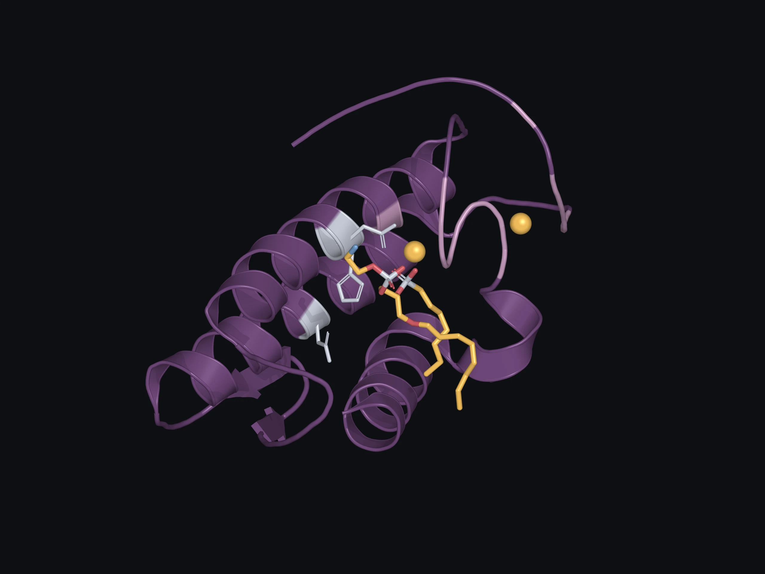
For secretory PLA₂ enzymes, the reaction begins at the boundary between water and a lipid surface. The enzyme binds to that surface, and a phospholipid enters a hydrophobic channel leading to the active site. The channel accommodates the lipid’s water-avoiding chains while positioning the region around the sn-2 bond ready for reaction.[3]
In the mechanism David L. Scott and colleagues proposed from structures of cobra- and bee-venom enzymes, a bound calcium ion within the active site helps position the substrate and stabilise the charged intermediate formed during hydrolysis. A histidine residue activates a water molecule for attack on the ester bond. The protein brings the reacting groups into an arrangement that supports cleavage, after which the fatty acid and lysophospholipid are released.[3]
A year later, the same group resolved human group IIA PLA₂, an enzyme better known from the synovial fluid of inflamed joints than from human skin, with a transition-state analogue bound in its active site. This inhibitor mimics aspects of the short-lived arrangement formed during hydrolysis, allowing us to examine how the enzyme accommodates it.[4] Comparing structures with and without it showed the enzyme’s hydrophobic channel changing shape as the analogue bound, a feat the authors found unique to this human enzyme.[4] The structure captures the bound analogue rather than a natural phospholipid being cut.
In this context, ‘free’ is chemistry shorthand for released from an ester, but it is not free in the literal sense. A free fatty acid’s long hydrocarbon chain still gives it a strong association with the lipid environment. What it has gained is a different chemical form, with a group that can now react.
The released fatty acid now contributes to the mixture twice over, through its chain and through its carboxyl group. Each leads to a different function: the chain into the organisation of the barrier, and the carboxyl group into its acid-base behaviour.
A fatty acid among other lipids
Once released, the carboxyl group can trade a proton with its surroundings. With the proton attached, it is written COOH. After giving it up, the group becomes the negatively charged carboxylate, COO⁻. The two forms exist in an equilibrium whose balance depends on pH and the molecular environment.
That second factor is easy to overlook. A carboxyl group packed among neighbouring lipids doesn’t necessarily behave as it would in dilute water. The measure of that behaviour is the pKₐ, the pH at which half of an acid’s molecules have given up their proton, so a higher value means a more reluctant acid. Renate Lieckfeldt and colleagues examined this in hydrated model mixtures of stratum corneum lipids in 1995. The fatty acids’ apparent pKₐ values ranged from 6.2 to 7.3, rising with the proportion of unsaturated oleic acid, and were 1.5 to 3 units higher than those of fatty acids in molecular solution.[5] Put plainly, packed among other lipids, a fatty acid holds on to its proton more tightly than it would in water.
Model lipid mixtures. The ionisation and packing experiments used hydrated lipid mixtures outside living tissue. They let researchers examine how the lipids behave as composition, pH or temperature changes.[5,6]
Below its pKₐ, the protonated form (COOH) predominates. From their model measurements, Lieckfeldt’s group estimated that about 90% of the fatty acids would be ionised at pH 7, while at a surface pH near 5 most would retain their proton.[5] The model results help explain why any account of a fatty acid’s ionisation has to include its surroundings. A pool that is mostly protonated still takes part in the equilibrium, exchanging protons with what surrounds it.
So cleavage makes an acid group available, but its contribution to tissue pH depends on the mixture around it. Other acids, ions and buffering groups are already present. It would be straightforward to count each cleavage as one proton added to a steadily growing pool, but the equilibrium doesn’t allow it.
The same surrounding lipids shape the other half of the fatty acid’s contribution, the physical half. Its chain interacts with neighbouring chains, affecting how closely they pack. Gooris and Bouwstra explored this using model membranes containing isolated human ceramides in 2007. At lower fatty-acid levels, orthorhombic and hexagonal packing coexisted; raising fatty acid to equimolar levels made the denser orthorhombic lattice dominant. Using deuterated fatty acids, they showed fatty acids and ceramides sharing that lattice, and extrapolated that the two share this domain in stratum corneum at 32 °C, skin temperature.[6] Packing is how a lipid mixture becomes a barrier, so a change in fatty-acid abundance is more than a change in inventory.
Acidity also changes how barrier lipids assemble, which brings the two halves of the story together. In Anna Nováčková’s 2021 study, multilamellar assembly of synthetic and extracted human stratum corneum lipids required acidic conditions. Neutral conditions changed their organisation and increased water permeability.[7] These systems contained no living epidermis, so the measurable change came from the lipids themselves, with no cell able to respond.
The chain becomes part of the material that forms the barrier, and the carboxyl group takes part in the acid-base chemistry around it. Interrupting its supply could therefore affect the tissue in more than one way, and the intervention experiments let us test that.
Interrupting the conversion
If the enzyme converts precursor into barrier material, blocking it should leave a gap. Peter Elias’s laboratory, with Mao-Qiang Man as first author, tested this in 1995 by inhibiting phospholipase activity after disrupting the barrier of hairless mice. Recovery slowed. Phospholipids accumulated, fatty-acid levels fell, and the extracellular membranes retained an immature appearance. Applying palmitic acid corrected the recovery defect, and incubating the treated stratum corneum with palmitic acid in vitro reversed the membrane changes. Within their experiments, an analogue that inhibits group II phospholipases but not group I left recovery unaffected, an early clue to which kind of enzyme was active.[1]
Mouse barrier recovery. This experiment tested recovery after acute barrier disruption. Palmitic-acid replacement restored recovery, while a separate incubation experiment examined the extracellular membrane changes.[1]
A question followed: did the damage come from the precursor piling up, or from the product being absent? The next result, in intact mouse skin, answered it. Repeated inhibition again impaired barrier function and reduced fatty acids, but this time the phospholipids did not pile up as they had before. Palmitic acid again corrected the abnormalities in this 1996 study.[8] Precursor had piled up in one experiment and not in the other, while the shortage of released fatty acids was common to both. The authors concluded that the barrier failed because of a lack of fatty acids, not because phospholipid accumulated.[8]
Replacement also depended on the fatty acid used. Linoleic acid did not reproduce the palmitic-acid rescue in these studies, and in 1995 neither did lysolecithin, the reaction’s other product.[1,8] The chain supplied therefore changes the outcome: adding an acid was not enough to guarantee the same result.
Yet the experiments left another possibility open. The missing fatty acids were no longer part of the lipid mixture, and the tissue had also lost their contribution to acid-base chemistry. How much of the resulting defect depended on that change in acidity?
Restoring acidity while fatty acids remain low
Joachim Fluhr’s group investigated this question in mouse skin in 2001. Two chemically unrelated inhibitors, making an effect peculiar to one compound less likely, raised surface pH and impaired permeability barrier function. The stratum corneum also became less cohesive, and less able to withstand repeated tape stripping, in which layers are peeled away with adhesive tape.[9]
Here, replacement with palmitic, stearic or linoleic acid, the last of which had not rescued in the earlier studies, reversed the abnormalities. That left a problem. A fatty acid supplies a chain and an acid group together, like a tablet containing two drugs: if the patient recovers, the tablet cannot say which drug did it. An acidic buffer is one of those two drugs on its own, acidity with no lipid, so the researchers tried that next. It restored cohesion and resistance to stripping, even though fatty-acid levels remained reduced.[9] For cohesion and integrity, acidity alone was enough.
Part of the damage therefore followed the pH change itself: three hours at neutral pH produced comparable abnormalities in intact stratum corneum. In the inhibitor-treated skin, electron micrographs showed why cohesion had failed: desmosomes, the protein junctions that hold corneocytes together, were breaking down early, and desmoglein 1, one of their structural proteins, was reduced.[9] What the rescue leaves open is how much of the permeability defect came from altered acidity and how much from the missing lipid material.
That is why phospholipid processing has consequences for both the supply of lipid and the acidity of the tissue. It supplies fatty acids to the extracellular mixture, and the acidity that comes with it is needed for the stratum corneum to hold together. The buffer experiment separates those consequences far enough to show that lipid depletion alone does not explain everything observed.
The pathway also contributes while acidity is first developing. Fluhr’s later neonatal rat experiments found secretory PLA₂ activity rising by 66% over the first five days after birth. Surface pH declined from 6.63 towards the adult value of about 5.9 over seven to eight days. Inhibiting secretory PLA₂ delayed this acidification, while inhibiting NHE1 partly blocked it.[10] Both routes contributed to postnatal acidification in that setting. Human skin also starts out near neutral at birth, and takes anywhere from days to months to reach the acidity observed in adults.[10]
The group IIA structure from earlier shows how one enzyme accommodates a lipid, but leaves another question: which phospholipase is doing the work?
Finding the enzymes
Secretory PLA₂ is a family. Its members differ in where they occur and how they interact with phospholipids. Calcium activates them at micromolar concentrations that vary with the substrate, while their activity on vesicles of different phospholipid composition varied by up to four orders of magnitude.[2] An enzyme’s presence is therefore only part of the explanation: the reaction also needs calcium and a lipid surface the enzyme can work on.
Was the enzyme family in human skin at all? In 2000, Juliette Mazereeuw-Hautier’s team detected secretory PLA₂ activity in human skin and tape strips, with pancreatic-type IB immunolabelling near the boundary between the granular layer and stratum corneum, while type II was undetectable.[11] ‘Pancreatic-type’ is a class name: the enzyme was detected in epidermis, and the pancreas was not involved. A wider survey by Ulrike Haas’s group in 2005 found different isoforms in different epidermal layers, with IB, IIF and X predominantly in the upper suprabasal layers, V and IID in the basal and spinous layers, and IIA only weakly expressed. Expression changed during differentiation and in psoriatic tissue.[12]
The shared reaction does not give every family member the same access to lipids. Ying H. Pan’s group resolved human group X PLA₂ and compared it with the group IIA enzyme. Group X was not activated by negatively charged interfaces, and worked just as well on zwitterionic surfaces, which carry both charges but no net charge, as on anionic ones. The authors predicted it would target the zwitterionic outer membranes of mammalian cells. Group X also had a wider entrance to its active site. Their structural and biochemical findings linked these differences to how the enzymes interact with lipid surfaces of different composition.[13]
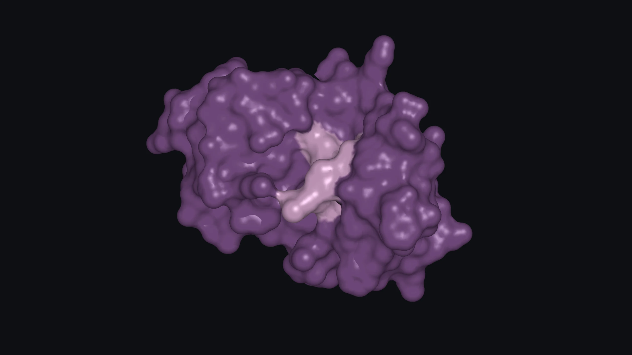
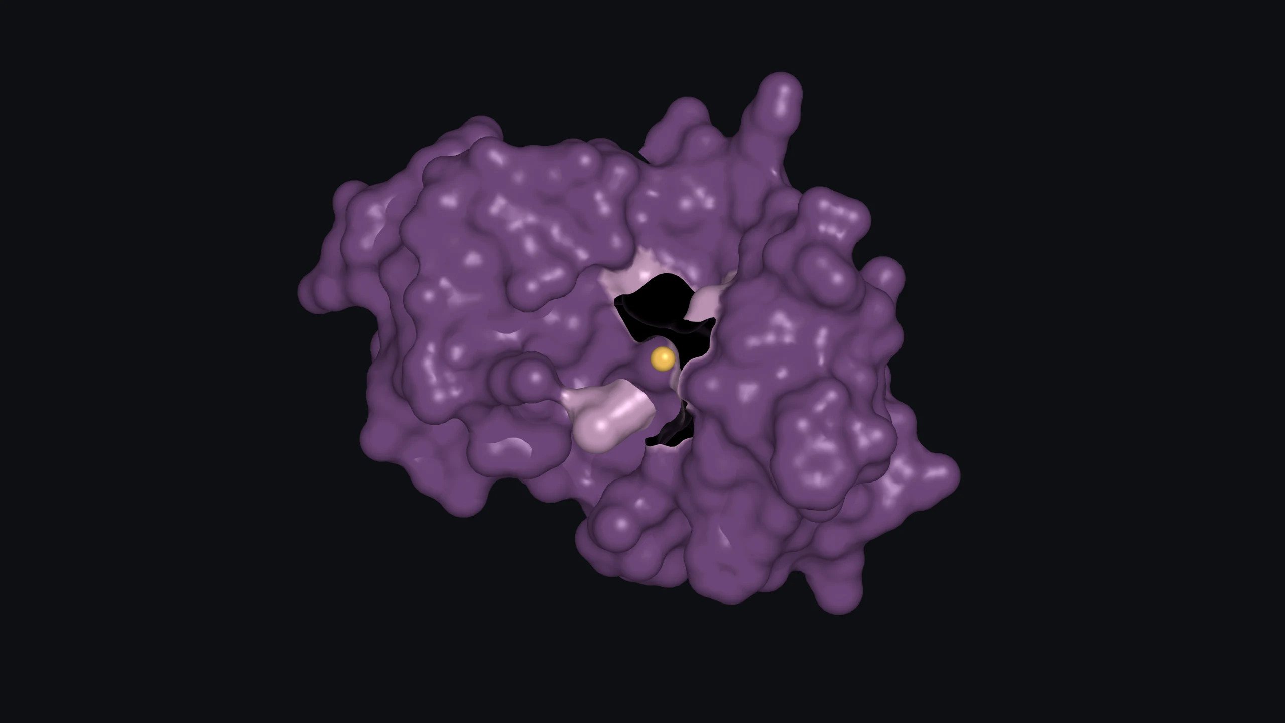
This brings the enzyme family’s variation down to the molecular surface: access depends on the protein and the lipids it encounters. The structures shown here are groups IIA and X, neither of them IIF, the isoform the genetic work below singles out. They explain features of catalytic recognition and lipid access; identifying the enzyme responsible for an effect in skin requires evidence from the tissue itself.
These findings show that the enzyme family is present in human epidermis, but they cannot say which member does what, and the older inhibitors could not either, as they do not single out one isoform. Genetics can narrow this down by removing one named enzyme at a time.
Group IIF has been tested in that way. In 2014, a group including Dusko Ilic and Theodora Mauro, both familiar from the NHE1 story, reported higher flank surface pH and slower early barrier recovery in mice lacking the enzyme.[14] They had chosen IIF because it was the most abundant isoform in mouse epidermis and, unlike IIA, rose with the nuclear-receptor agonists that speed acidification.[14] A year later, Kei Yamamoto’s group found that the picture was less uniform: the phenotype varied across the skin. Adult knockout mice had increased pH and water loss in abdominal tissue, accompanied by reduced fatty acids and ceramides, while dorsal skin did not show those changes. Among the secreted PLA₂ knockouts they compared, only loss of IIF raised water loss.[15]
The effect was restricted to abdominal skin, complicating a direct acid-supply explanation. The knockout stratum corneum was fragile even where it looked normal: corneocytes from back skin collapsed faster under sonication. Yamamoto’s group proposed that environmental stress, including friction against the cage floor, exposed that vulnerability. The abdomen is, after all, the side of a mouse that is more exposed to contact with the cage floor. In their interpretation, barrier disruption led to the abdominal changes in pH and lipid levels. Friction was not directly tested as the cause.[15]
The knockouts were also strikingly protected from experimental psoriasis, contact dermatitis and skin cancer, while mice overexpressing IIF developed psoriasis-like hyperplasia.[15] In the same study, a lipid product called a lysoplasmalogen restored aspects of activation in keratinocytes lacking IIF, both in culture and in the mice.[15] A lysoplasmalogen is what remains of the substrate once its fatty acid has been released, much as the lysophospholipid remains in the conventional case described earlier. Its effects give the cleavage a second job: signalling as well as a supply of fatty acids.
Removing IIF alters signalling and barrier stability as well as lipid composition, so the pH change can’t be read as a direct acidifying role. Two decades after the first inhibitor experiments, the answer to which phospholipase is doing the work is, for now, a family: the evidence supports the group more firmly than any single member.
Maintaining acidity as the tissue changes
A pathway that contributes acidity should be adjustable, so the next question is how the epidermis regulates it. Enzyme activity changes during development, and researchers have also altered it experimentally through signals that regulate lipid metabolism.
In 2009, Fluhr’s group came back to neonatal rats with the opposite experiment. Instead of blocking the pathway, they tried to speed up acidification, treating the animals with compounds activating PPARα, a nuclear receptor that switches on genes involved in lipid metabolism. Acidification accelerated together with increased secretory PLA₂ activity, and a broad phospholipase inhibitor blocked the additional acidifying effect; NHE1 staining didn’t change, although the authors noted that the inhibitor, bromophenacyl bromide, acts on more than PLA₂.[16] This supports a regulated contribution from the pathway, within the wider changes in differentiation and lipid metabolism produced by PPARα activation. Ilic’s group later found that PPARα agonists raised IIF but not IIA in mouse epidermis, which ties this result to the isoform the knockouts implicated.[14]
Compensation brings us back to filaggrin. In Kateřina Vávrová’s study of reconstructed human skin, a laboratory-grown model of epidermis, reducing filaggrin lowered its acidic breakdown products, PCA and urocanic acid, yet surface pH remained acidic. NHE1 was upregulated first, and by day 14 IIA expression and secretory PLA₂ activity had increased. Ceramide processing was unaffected, but free fatty acids doubled, and the authors linked that excess to less ordered lipid lamellae and higher permeability.[17] Fluhr, by then in Berlin, was among its authors.[17]
Reconstructed human skin. Filaggrin was reduced in laboratory-grown epidermis. The study followed acidification, enzyme expression, lipid composition and permeability as the constructs developed.[17]
So the tissue held its acidity while the lipid barrier still changed. In this model the fix carried its own cost: the phospholipase activity that kept the surface acidic also produced the surplus fatty acid the authors linked to disordered lamellae. The investigators described this as compensation, although what initiated the response remained unresolved.
So the tissue held its acidity while the lipid barrier still changed.
At the boundary with the stratum corneum, secretion begins a conversion that continues outside the cell. A fatty acid released there becomes part of the developing lipid barrier, with chemistry it could not express while esterified. Part of the epidermis’s acidic environment emerges through that conversion. How large that part is, and which enzymes carry it, is still being worked out.
Glycerophospholipid. A lipid with a glycerol backbone and a phosphate-containing head group. In the conventional form described here, two fatty-acid chains are attached through ester bonds.
sn-2. The second position on the glycerol backbone. PLA₂ cleaves the ester bond linking a fatty-acid chain at this position.
Lysophospholipid. The lipid left after one fatty-acid chain is removed from a conventional glycerophospholipid. It retains its head group and the remaining chain.
pKₐ. The pH at which the protonated and deprotonated forms of an acid are present in equal proportions. In a lipid mixture the measured value is an apparent pKₐ, because it describes that particular molecular environment.
Orthorhombic. A dense, ordered arrangement of neighbouring lipid chains, also called orthorhombic packing. The packing studies examine how fatty acids and ceramides share this arrangement.
Zwitterionic. Carrying both positive and negative charges while having no overall electrical charge. This describes the lipid head groups discussed in the group X comparison.
Lysoplasmalogen. The product left when PLA₂ removes the fatty-acid chain at sn-2 from a plasmalogen. It retains the ether-linked chain at sn-1. The IIF experiments examined an ethanolamine lysoplasmalogen.
PPARα. Peroxisome proliferator-activated receptor alpha, a nuclear receptor that regulates gene expression involved in lipid metabolism and epidermal differentiation.
References
1. Mao-Qiang, M., Feingold, K.R., Jain, M., et al. (1995). Extracellular processing of phospholipids is required for permeability barrier homeostasis. Journal of Lipid Research, 36(9), pp. 1925-1935. https://doi.org/10.1016/S0022-2275(20)41111-3
2. Singer, A.G., Ghomashchi, F., Le Calvez, C., et al. (2002). Interfacial kinetic and binding properties of the complete set of human and mouse groups I, II, V, X, and XII secreted phospholipases A₂. Journal of Biological Chemistry, 277(50), pp. 48535-48549. https://doi.org/10.1074/jbc.M205855200
3. Scott, D.L., White, S.P., Otwinowski, Z., et al. (1990). Interfacial catalysis: the mechanism of phospholipase A₂. Science, 250(4987), pp. 1541-1546. https://doi.org/10.1126/science.2274785
4. Scott, D.L., White, S.P., Browning, J.L., et al. (1991). Structures of free and inhibited human secretory phospholipase A₂ from inflammatory exudate. Science, 254(5034), pp. 1007-1010. https://doi.org/10.1126/science.1948070. Structure record: https://www.rcsb.org/structure/1POE
5. Lieckfeldt, R., Villalaín, J., Gómez-Fernández, J.C., et al. (1995). Apparent pKₐ of the fatty acids within ordered mixtures of model human stratum corneum lipids. Pharmaceutical Research, 12(11), pp. 1614-1617. https://doi.org/10.1023/A:1016280714593
6. Gooris, G.S. and Bouwstra, J.A. (2007). Infrared spectroscopic study of stratum corneum model membranes prepared from human ceramides, cholesterol, and fatty acids. Biophysical Journal, 92(8), pp. 2785-2795. https://doi.org/10.1529/biophysj.106.094292
7. Nováčková, A., Sagrafena, I., Pullmannová, P., et al. (2021). Acidic pH is required for the multilamellar assembly of skin barrier lipids in vitro. Journal of Investigative Dermatology, 141(8), pp. 1915-1921.e4. https://doi.org/10.1016/j.jid.2021.02.014
8. Mao-Qiang, M., Jain, M., Feingold, K.R., et al. (1996). Secretory phospholipase A₂ activity is required for permeability barrier homeostasis. Journal of Investigative Dermatology, 106(1), pp. 57-63. https://doi.org/10.1111/1523-1747.ep12327246
9. Fluhr, J.W., Kao, J., Jain, M., et al. (2001). Generation of free fatty acids from phospholipids regulates stratum corneum acidification and integrity. Journal of Investigative Dermatology, 117(1), pp. 44-51. https://doi.org/10.1046/j.0022-202x.2001.01399.x
10. Fluhr, J.W., Behne, M.J., Brown, B.E., et al. (2004). Stratum corneum acidification in neonatal skin: secretory phospholipase A₂ and the sodium/hydrogen antiporter-1 acidify neonatal rat stratum corneum. Journal of Investigative Dermatology, 122(2), pp. 320-329. https://doi.org/10.1046/j.0022-202X.2003.00204.x
11. Mazereeuw-Hautier, J., Redoules, D., Tarroux, R., et al. (2000). Identification of pancreatic type I secreted phospholipase A₂ in human epidermis and its determination by tape stripping. British Journal of Dermatology, 142(3), pp. 424-431. https://doi.org/10.1046/j.1365-2133.2000.03351.x
12. Haas, U., Podda, M., Behne, M., et al. (2005). Characterization and differentiation-dependent regulation of secreted phospholipases A₂ in human keratinocytes and in healthy and psoriatic human skin. Journal of Investigative Dermatology, 124(1), pp. 204-211. https://doi.org/10.1111/j.0022-202X.2004.23513.x
13. Pan, Y.H., Yu, B.-Z., Singer, A.G., et al. (2002). Crystal structure of human group X secreted phospholipase A₂. Electrostatically neutral interfacial surface targets zwitterionic membranes. Journal of Biological Chemistry, 277(32), pp. 29086-29093. https://doi.org/10.1074/jbc.M202531200. Structure record: https://www.rcsb.org/structure/1LE6
14. Ilic, D., Bollinger, J.M., Gelb, M., et al. (2014). sPLA₂ and the epidermal barrier. Biochimica et Biophysica Acta, 1841(3), pp. 416-421. https://doi.org/10.1016/j.bbalip.2013.11.002
15. Yamamoto, K., Miki, Y., Sato, M., et al. (2015). The role of group IIF-secreted phospholipase A₂ in epidermal homeostasis and hyperplasia. Journal of Experimental Medicine, 212(11), pp. 1901-1919. https://doi.org/10.1084/jem.20141904
16. Fluhr, J.W., Man, M.-Q., Hachem, J.-P., et al. (2009). Topical peroxisome proliferator activated receptor activators accelerate postnatal stratum corneum acidification. Journal of Investigative Dermatology, 129(2), pp. 365-374. https://doi.org/10.1038/jid.2008.218
17. Vávrová, K., Henkes, D., Strüver, K., et al. (2014). Filaggrin deficiency leads to impaired lipid profile and altered acidification pathways in a 3D skin construct. Journal of Investigative Dermatology, 134(3), pp. 746-753. https://doi.org/10.1038/jid.2013.402
18. Miki, Y., Higashisaka, N., Inubushi, H., et al. (2026). PLA2G2F/lysoplasmalogen axis links epidermal lipid metabolism to type 2 inflammation and itch in atopic dermatitis. Frontiers in Immunology, 17, 1880255. https://doi.org/10.3389/fimmu.2026.1880255
A short note
The intervention studies most directly linking phospholipid processing to acidity were performed in rodents. Human tissue and reconstructed epidermis support biological relevance, while the contribution of individual enzymes to acidity in intact adult human skin remains unresolved. The lipid-assembly experiments used model systems.[6,7,11,12,14,15,17]
The older inhibitors do not selectively identify one isoform. Fatty-acid rescue shows that replacement overcomes a defect under the tested conditions; it does not identify the normal source of every barrier fatty acid. Linoleic-acid rescue differed between the earlier processing studies and the 2001 acidification study. Their interventions and endpoints differed, and the reason for the different outcomes remains unresolved.[1,8,9] Surface-pH changes also do not allocate a fixed share of acid production to either phospholipases or NHE1.[10]
Ilic’s 2014 paper combines a review with original experiments.[14] Yamamoto’s substrate work linked IIF to DHA-containing ethanolamine plasmalogens and lysoplasmalogen products.[15] More recent research connects the PLA2G2F/lysoplasmalogen pathway to inflammatory signalling and itch in experimental atopic dermatitis, with human lipid measurements associated with disease severity.[18] That work addresses signalling rather than establishing another acidifying contribution.
Mazereeuw-Hautier’s human study included Institut de Recherche Pierre Fabre affiliations.[11] Miki’s 2026 study reports public funding as well as support from Kose Cosmetology, Ono Medical Research and Terumo Life Science foundations.[18] This article was written independently, without sponsorship, affiliate links or product recommendations.