
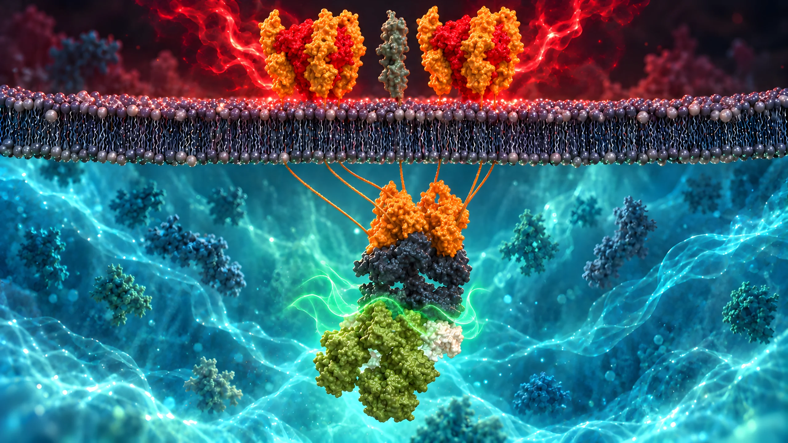
In the previous article, caspase-8 appeared where we didn’t expect it: keratinocytes beginning to differentiate. Inhibiting it interfered with their commitment, although those cells were not undergoing ordinary apoptosis. A cell on its way to becoming the barrier appeared to need a death protein to get there. That left a question.
Caspase-8 also belongs to the machinery that death receptors recruit at the cell surface, so the protein a keratinocyte seems to use for differentiation is the same one a death receptor uses to start killing it. When one of those receptors receives a signal, what prevents a living keratinocyte from dying before it has finished building the epidermis?
Staining patterns in inflamed human skin offered a clue. In acute eczema, activated T cells enter the tissue and some keratinocytes undergo apoptosis. Nicole Armbruster’s group stained inflamed skin for cleaved caspase-3, which marks cells dying by apoptosis, and found the dying cells predominantly above the basal layer[1]. Then they stained for cFLIP, a protein we will soon meet, and found the reverse. Its expression was highly restricted to the basal keratinocytes, the layer where the dying cells were far less common. The basal layer contains the cells that continually replenish the epidermis, so the pattern had a logic to it: the layer that has to persist was the layer carrying cFLIP.
The group then tested whether cFLIP could do the protecting. When the researchers transduced a vector expressing the long isoform, cFLIP-L, into HaCaT keratinocytes and cultured them with activated CD4 T cells, those cells were effectively protected from T-cell-mediated apoptosis.[1]
It was a good clue, but an incomplete one. The pattern suggested that basal cells carried some form of protection that their suprabasal neighbours lacked. The staining was a correlation in tissue; the protection was a gain of function in a cell line. Neither showed that cFLIP alone explains where death occurred in eczema, and neither showed what happens to skin that loses it. But it did make an interesting experimental question. What would happen to an epidermis whose keratinocytes lost cFLIP?
Three versions of the same protein
cFLIP was characterised in 1997 as an inhibitor of death receptor signalling: it could block apoptosis induced by every human death receptor then known. Its name, cellular FLICE-inhibitory protein, is derived from that first reputation; FLICE was an earlier name for caspase-8.[2] I first met cFLIP in cancer biology, where the aim was to block its activity: my master’s research asked whether a novel cFLIP inhibitor could sensitise breast cancer stem-like cells to TRAIL-mediated death. In skin the question runs the opposite direction. Here the cells need it.
Human cells translate three isoforms. A long form, cFLIP-L, of 480 amino acids; a short form, cFLIP-S, of 221; and a third short form named for the Raji cell line it was found in, cFLIP-R, of 213.[2,3]
All three isoforms begin with two death effector domains, which allow cFLIP to join the complex assembling beneath an activated death receptor. What follows them separates the isoforms. cFLIP-L contains a caspase-like domain; it is structurally homologous to caspase-8’s, but the active-site cysteine has been substituted by a tyrosine. cFLIP-L is therefore catalytically inactive and cannot cut. cFLIP-S and cFLIP-R terminate after the death effector domains and contain no caspase-like domain at all.[2,3]
Take note of cFLIP-L. A caspase-8 lookalike that cannot cut sounds like a dead end, but the next result is the first sign that this is not the case.
Early accounts treated the regulation as competition. cFLIP and caspase-8 bind the adaptor protein FADD through the same type of domain, so whichever is more abundant should win the site. Michelle Hughes and colleagues used a functional reconstituted death-inducing signalling complex (DISC) and the prediction failed in both directions: cFLIP did not displace procaspase-8 from FADD, and without procaspase-8 present, almost no cFLIP was recruited.[4]
The order runs opposite to the one initially assumed. FADD recruits procaspase-8 first; procaspase-8 then recruits cFLIP and heterodimerises with it. Which heterodimer forms depends on the ratio of unbound cFLIP to procaspase-8, and the two have opposite effects. A procaspase-8:cFLIP-L pair exhibits localised enzymatic activity and acts preferentially as an activator, promoting the recruitment of further procaspase-8 molecules. A procaspase-8:cFLIP-S pair lacks activity and potently blocks procaspase-8 activation.[4] cFLIP-L also inhibits caspase-8 activation when present at higher concentrations. Its effect depends on its abundance relative to procaspase-8.[4]
Some of this was established in keratinocytes. The team purified CD95 (Fas) DISCs from HaCaT cells engineered to stably overexpress cFLIP-S. In those cells, CD95 ligand no longer triggered caspase-8 cleavage or apoptosis. In control DISCs, caspase-8 was about six times as abundant as FADD, consistent with several procaspase-8 molecules assembling through each FADD. With excess cFLIP-S, FADD recruitment remained similar, but FADD, caspase-8 and cFLIP were present in roughly equal amounts. cFLIP-S had not kept caspase-8 out of the DISC. It had limited how much more could assemble there, preventing the interactions needed to activate it.[4]
So cFLIP isn’t a binary switch held over caspase-8. It behaves more like a dial: its form and its abundance set how caspase-8 behaves, and with it which route to death remains available.[4]
Remove the safeguard
How the deletion worked. The mice carried two floxed cFLIP alleles and tamoxifen-responsive Cre under the keratin 14 promoter. Five daily applications of tamoxifen to depilated adult skin activated Cre in the treated epidermis, where it excised cFLIP. Controls also received tamoxifen, but carried wild-type cFLIP alleles. The team checked excision by multiplex PCR.[5]
The obvious experiment was to delete cFLIP from the keratinocytes entirely, but the first attempt at it largely defeated itself. Diana Panayotova-Dimitrova’s team bred mice to lose cFLIP in the epidermis, yet the experiment had a high rate of embryonic loss between days 11 and 14 (60%); the rare pups born still retained epidermal cFLIP. The authors suggested that the survivors retained cFLIP because Cre, the enzyme that carries out the deletion, had been silenced, or because cells that lost cFLIP failed to persist.[5]
So, they changed the question. Instead of deleting cFLIP from birth and recording the phenotype there, the researchers used a system in which they could trigger its removal from a restricted area of adult skin and watch an established epidermis respond over the following days.[5] These deletion experiments removed all cFLIP isoforms rather than isolating the contribution of an individual.
At 48 hours, cFLIP staining had fallen, particularly in basal keratinocytes. Looking at the tissue as a whole, though, there was little obvious change; the layers still appeared organised. Keratin 14, keratin 10 and loricrin, proteins used to follow different stages of epidermal differentiation, retained their expected staining patterns. Yet some keratinocytes already showed active caspase-3, one of the executioner enzymes of apoptosis. These first caspase-3-positive cells outnumbered both the dyskeratotic (abnormally keratinised) cells and the keratinocytes positive for TUNEL, a test that detects DNA fragmentation. Every dyskeratotic cell examined also stained for cleaved caspase-3. Electron microscopy also found the condensed chromatin and cell shrinkage associated with this form of death.[5]
The death machinery was active before the epidermis visibly lost its organisation.
After topical tamoxifen | adult mouse skin
48 hours
cFLIP staining has fallen, especially in basal cells.
Death | cleaved caspase-3 is detectable.
Architecture | layers and K14, K10 and loricrin patterns still appear organised.
72 hours
cFLIP is absent from the epidermis.
Death | dyskeratotic keratinocytes are frequent.
Architecture | thickening, inflammation and disturbed marker patterns are evident.
96 hours
Architecture | large areas of epidermis disintegrate.
Lesions | neutrophil-filled pustules and subepidermal blisters develop.
The times run from the start of tamoxifen treatment. The panels show observed changes, not a measured severity scale.
By 72 hours the effect was difficult to miss. The treated skin was red and thickened, inflammatory cells had entered, and dyskeratotic keratinocytes had become frequent. By 96 hours the epidermis had disintegrated across large regions, with pustules full of neutrophils and blisters beneath. The authors describe the response as biphasic: an inflammatory phase first, then the wave of cell loss that took the full thickness of the epidermis. Daily topical disinfection in a separate experiment changed neither the timing nor the severity of the disease, arguing against bacterial overgrowth as the event that started it.[5]
One working copy of Cflar, the gene encoding cFLIP, was enough. Mice carrying a single wild-type Cflar allele showed no spontaneous apoptosis or caspase activation in the epidermis; losing both copies exposed the epidermis to apoptosis.[5]
Removing cFLIP had damaged an established epidermis. What the experiment had not yet identified was the pathway that made its loss so consequential.
Which signal was getting through?
A second way to delete cFLIP. Tamoxifen produced incomplete deletion in primary keratinocyte cultures, so the team delivered Cre with a lentiviral vector. This allowed them to examine spontaneous death after more complete deletion and compare a TNF-binding reagent with Fas ligand and TRAIL decoy receptors. TNF-R2-Fc bound the ligand; its name does not mean that TNFR2 on the keratinocytes caused the death.[5]
The researchers turned to primary mouse keratinocytes, which let them observe the cells alone. Removing cFLIP left them, in the authors’ word, exquisitely sensitive to added Fas ligand or tumour necrosis factor. The tamoxifen system deleted the gene only partly in culture, so the researchers delivered Cre by virus instead. More revealing, with complete cFLIP deletion, between 50% and 70% of the cells died by day 4, with no death ligand added at all.[5]
Nothing had been added to kill them, so something in the dish was. One possible explanation was that the cells were supplying a signal of their own. cFLIP-depleted keratinocytes increased TNF production. A soluble receptor protein that bound TNF protected the cultures from their spontaneous death. Blocking Fas ligand or TRAIL did not. The TNF rise was a response to losing cFLIP rather than to the virus or the enzyme it carried, which the researchers checked separately. In this case, TNF had become part of a self-sustaining, autocrine route into apoptosis: keratinocytes produced it, then responded to it in a way cFLIP would normally restrain.[5]
TNF is often discussed as though it issues a single death instruction, but binding to TNFR1 does not automatically kill a cell. The first signalling complex formed at the receptor supports inflammatory and survival responses. Under conditions that permit a death response, a secondary complex containing proteins such as FADD and caspase-8 forms within the cell. cFLIP participates at this point. Remove it, and TNF signalling has a much easier route into caspase-dependent apoptosis.[5,6]
So, would intercepting TNF protect the skin of the mice as well? Pretreatment with a soluble TNF-binding receptor significantly reduced the keratinocytes staining for active caspase-3 at 72 hours. Later, TNF blockade also reduced visible skin damage at 96 and 120 hours, but did not prevent it. At later time points, the visible difference between the TNF-blocked mice and the IgG-treated controls became less pronounced. TNF had driven early keratinocyte apoptosis, but blocking it did not stop the skin disease.[5] Here, TNF was not the whole explanation.
The rescue that stopped short
A later study gave TNF a more decisive test. Feoktistova’s group again triggered cFLIP deletion in the adult epidermis. This time, they did so in mice that also lacked TNF throughout the body. If TNF caused everything that followed cFLIP loss, removing both should have restored the skin. [6]
It made a striking difference. Weight loss and mortality were completely rescued by the absence of TNF, compared with TNF-producing controls. Skin disease developed later and was milder, and the skin recovered faster: active caspase-3 fell significantly at 72 hours, skin thickening was significantly reduced. Weight loss and mortality turned out to be TNF-dependent; the skin disease only partly so.[6]
Same adult epidermal deletion | different TNF backgrounds
Local epidermal cFLIP lossTNF present
Severe disease
Keratinocyte apoptosis and skin inflammation followed deletion. Mice lost weight and died.
Systemic outcome | weight loss and mortality
Local epidermal cFLIP lossTNF absent throughout the animal
Rescue that stopped short
Weight loss and mortality were rescued. Epidermal apoptosis fell, and skin disease was delayed and milder, but inflammation and disrupted differentiation markers remained.
Systemic outcome rescued | skin partly improved
The whole-animal Tnf knockout is a different test from treating mice with the soluble TNF-binding reagent.
Inflammation still appeared. Keratin 14, keratin 10 and loricrin staining remained disturbed, showing that the organisation of differentiating keratinocytes was still affected.[6]
The inflammatory cells told an even more specific story. TNF loss reduced the arrival of Gr-1-positive cells, a population including granulocytes. It did not reduce the number of infiltrating CD3-positive T cells at the early time point examined. There was therefore no single inflammatory response for TNF to switch off. Some parts receded while others continued.[6] The array gave a further insight. Genes associated with epidermal barrier function, among them kallikrein-related peptidase 8, filaggrin, IL-18 and caspase-14, were downregulated in a TNF-dependent way, which the authors offer as part of why the TNF-deficient skin looked better.[6]
Taking the receptor away
A different genetic test. Piao’s mice used Krt5-Cre to delete floxed cFLIP during epidermal development, alongside a whole-animal TNFR1 knockout (Tnfrsf1a−/−). There was no topical tamoxifen step. This tested which death routes remained without TNFR1 during development, under different conditions from the adult TNF-knockout experiment.[7]
If TNF itself only accounted for part of the damage, the rest had to come from somewhere else. In a separate study, Xuehua Piao’s team went upstream of the cytokine to its receptor. They deleted cFLIP from the developing epidermis and, instead of removing TNF, removed TNFR1, the receptor responsible for the TNF death pathway examined here.[7]
A full TNFR1 knockout was required for the pups to reach birth. Mice keeping a single functional TNFR1 allele died in utero of massive keratinocyte apoptosis. Those lacking TNFR1 entirely were born at the expected Mendelian ratio and looked well, though some basal cells were already irregularly arranged. Over the coming first week dermatitis appeared, water loss through the skin increased, keratinocytes died by apoptosis, and the pups died soon afterwards.[7]
Fas ligand and TRAIL each killed cFLIP-deficient keratinocytes in culture. Blocking both ligands together in the pups substantially prolonged survival. This rescue also stopped short: the treated pups still developed severe dermatitis and died at around ten days.[7]
Looking at the rescues discussed here and a pattern appears. Blocking TNF lessened the early damage in adult mice. Removing it completely saved the animals’ weight and survival, but not their skin. Removing the receptor carried the pups to birth, but they died soon afterwards. Blocking Fas ligand and TRAIL as well extended survival to around ten days. Each removal bought time. None bought a cure. The models differ, so this remains a sequence of comparisons rather than a single experiment.[5,6,7]
The skin showed where the damage landed. Inflammatory cytokines rose, and epidermal differentiation markers, loricrin, filaggrin and keratin 10, were lost; keratin 5 in the basal layer was untouched. Treating primary keratinocytes with IL-6, and to a lesser extent IL-17A, reproduced that loss. IL-6 also activated STAT3, which the authors proposed as part of the route to reduced differentiation-marker expression.[7] The authors concluded that cFLIP protects differentiation by preventing keratinocyte death and the inflammatory cytokine response that follows. Cytokines such as IL-6 then suppress differentiation in the surviving cells.[7] Blocking IL-6 in the pups slightly prolonged survival but did not prevent the lethal disease.[7]
The disturbed differentiation markers need a closer look. At 48 hours, cFLIP staining had fallen, particularly in basal keratinocytes, and apoptosis was already detectable while differentiation markers remained normally arranged. By 72 hours, the epidermis fully lacked cFLIP expression and the differentiation-marker patterns were disturbed. Their later disruption occurred in a tissue that was dying and inflamed; therefore, it does not on its own establish a direct role for cFLIP in differentiation. The disruption also persisted in the TNF-deficient mice so whatever disturbs differentiation here is not only TNF-driven death.[5,6]
The developmental study does not identify every signal responsible for the residual disease in Feoktistova’s adult mice. It shows why a TNF rescue doesn’t need to settle the story. cFLIP stands at signalling machinery used by several death receptor pathways. Protecting a keratinocyte against one ligand leaves it vulnerable to others in its environment.[7]
Why not remove caspase-8 as well?
If cFLIP restrains a death route involving caspase-8, surely removing caspase-8 should make keratinocytes safer?
Ricardo Weinlich’s group removed either cFLIP or caspase-8 from local areas of adult mouse skin. Both deletions produced the same picture: thickened, inflamed skin, infiltrating immune cells and staining revealed dying keratinocytes in both. Still, appearance alone offered little explanation for why the cells were dying.[8]
The researchers then removed RIPK3, a protein required for a different regulated route to cell death called necroptosis. In this route RIPK3 activates MLKL, which in turn damages the cell membrane. Removing RIPK3 rescued the caspase-8-deficient skin; minimal epidermal hyperplasia, rare dead keratinocytes and minimal dermal inflammation were observed. In the cFLIP-deficient skin it neither prevented nor lessened the disease.[8]
Adult skin | test the route by removing RIPK3
Remove caspase-8Skin injury
RIPK3 removal rescues
With RIPK3 also absent, little epidermal hyperplasia, keratinocyte death or dermal inflammation remained.
RIPK3-dependent death
Remove cFLIPSkin injury
RIPK3 removal does not rescue
The skin disease persisted. Dying keratinocytes in the cFLIP-deficient tissue stained for cleaved caspase-3.
Apoptotic death
The genetic rescue distinguishes the outcomes. TUNEL staining on its own does not identify necroptosis.
The two deletions had not exposed the same death. Where cFLIP was removed, the dying keratinocytes stained for cleaved caspase-3; the authors noted the death as apoptosis. Where caspase-8 was singularly removed the death ran through RIPK3, and the evidence for that is the rescue itself: deleting caspase-8 in mice already lacking RIPK3 largely prevented keratinocyte death.[8] TNF appeared in the skin after either deletion, and neutralising it protected against the effects of both. The authors placed TNF upstream of two different outcomes: apoptosis after cFLIP loss and RIPK3-dependent damage after caspase-8 loss.[8]
Earlier experiments help explain why. The activity retained by the caspase-8–cFLIP-L pair is important here: it suppresses RIPK3-dependent death without committing the cell to apoptosis.[9] The same partnership restrains the caspase-8 activity that would drive apoptosis. Remove cFLIP and both restraints are lost. Consistent with this, the embryonic lethality of cFLIP loss is relieved only when both RIPK3 and FADD are absent.[8,9]
Too little control over caspase-8 exposes apoptosis. Remove caspase-8 itself, and a separate danger is no longer held in check. The cell needs the machinery, but it also needs to govern its use. If caspase-8 is the devil in this story, cFLIP is not trying to banish it. The cell cannot do without the machinery, so it dances with it, and the dance is how a dangerous partner is held at bay.[8,9]
The point reaches back to the previous article. There, caspase activity appeared during commitment to differentiation without making that commitment ordinary apoptosis. Here, removing one of its regulators exposed how forcefully the same wider machinery could damage living epidermis. A caspase’s presence, and even some of its activity, does not by itself tell us how the cell’s story will end.[5,8,9]
Back to human skin
In healthy human epidermis, Panayotova-Dimitrova’s group found strong cFLIP staining in the basal layer and no detectable staining for cleaved caspase-8. Lesions from Stevens–Johnson syndrome and toxic epidermal necrolysis showed reduced cFLIP staining in areas with increased cleaved caspase-8 staining.[5]
The lesion samples cannot tell us whether reduced cFLIP preceded cell death or resulted from tissue injury. They do return us to Armbruster’s observation: cFLIP was strongest in basal keratinocytes, while apoptosis appeared mainly above them.[1,5]
A keratinocyte must survive long enough to help build a barrier whose outermost cells are no longer alive. That survival requires active control of its death machinery. cFLIP restrains apoptosis while preserving a caspase-8 partnership that suppresses another route to death. Remove either partner and living epidermis breaks down. After all, the cells that build the barrier need protection of their own.[8,9]
Apoptosis. Regulated cell death in which caspases dismantle the cell. Condensed chromatin and cell shrinkage are characteristic signs.
Necroptosis. A regulated route to death in which RIPK3 activates MLKL, leading to damage of the cell membrane.
DISC. The death-inducing signalling complex assembled at activated CD95 (Fas). FADD, procaspase-8 and cFLIP are among its components.
Death effector domain. A protein-binding region shared by FADD, procaspase-8 and cFLIP that helps organise their assembly.
Heterodimer. A pair of different proteins. Here, procaspase-8 pairs with a cFLIP isoform.
Autocrine signalling. Cells responding to a signal they also produce. In culture, cFLIP-depleted keratinocytes produced TNF that contributed to their own death.
Floxed allele. A gene copy flanked by loxP DNA sites, allowing Cre recombinase to excise it.
CreER. A tamoxifen-responsive form of Cre recombinase. The keratin 14 promoter directs where it is expressed; tamoxifen controls when it acts.
TUNEL. A test for fragmented DNA. A positive result alone does not identify which route to cell death occurred.
References
1. Armbruster, N., Trautmann, A., Bröcker, E.-B., et al. (2009). Suprabasal spongiosis in acute eczematous dermatitis: cFLIP maintains resistance of basal keratinocytes to T-cell-mediated apoptosis. Journal of Investigative Dermatology, 129(7), pp. 1696–1702. https://doi.org/10.1038/jid.2008.438
2. Irmler, M., Thome, M., Hahne, M., et al. (1997). Inhibition of death receptor signals by cellular FLIP. Nature, 388(6638), pp. 190–195. https://doi.org/10.1038/40657
3. Golks, A., Brenner, D., Fritsch, C., et al. (2005). c-FLIPR, a new regulator of death receptor-induced apoptosis. Journal of Biological Chemistry, 280(15), pp. 14507–14513. https://doi.org/10.1074/jbc.M414425200
4. Hughes, M.A., Powley, I.R., Jukes-Jones, R., et al. (2016). Co-operative and hierarchical binding of c-FLIP and caspase-8: a unified model defines how c-FLIP isoforms differentially control cell fate. Molecular Cell, 61(6), pp. 834–849. https://doi.org/10.1016/j.molcel.2016.02.023
5. Panayotova-Dimitrova, D., Feoktistova, M., Ploesser, M., et al. (2013). cFLIP regulates skin homeostasis and protects against TNF-induced keratinocyte apoptosis. Cell Reports, 5(2), pp. 397–408. https://doi.org/10.1016/j.celrep.2013.09.035
6. Feoktistova, M., Makarov, R., Leverkus, M., et al. (2020). TNF is partially required for cell-death-triggered skin inflammation upon acute loss of cFLIP. International Journal of Molecular Sciences, 21(22), 8859. https://doi.org/10.3390/ijms21228859
7. Piao, X., Miura, R., Miyake, S., et al. (2019). Blockade of TNFR1-dependent and TNFR1-independent cell death is crucial for normal epidermal differentiation. Journal of Allergy and Clinical Immunology, 143(1), pp. 213–228.e10. https://doi.org/10.1016/j.jaci.2018.02.043
8. Weinlich, R., Oberst, A., Dillon, C.P., et al. (2013). Protective roles for caspase-8 and cFLIP in adult homeostasis. Cell Reports, 5(2), pp. 340–348. https://doi.org/10.1016/j.celrep.2013.08.045
9. Dillon, C.P., Oberst, A., Weinlich, R., et al. (2012). Survival function of the FADD–caspase-8–cFLIP-L complex. Cell Reports, 1(5), pp. 401–407. https://doi.org/10.1016/j.celrep.2012.03.010
A short note
The principal deletion studies used genetically modified mice. The adult experiments triggered cFLIP loss in restricted, treated skin; the Piao study examined deletion during development alongside loss of TNFR1. Their different ages and genetic designs are reasons to use the latter study to identify an additional route into damage, rather than treating its outcome as a direct test of the remaining disease in the adult TNF-deficient mice.[5,6,7]
The primary mouse keratinocyte cultures established an important role for TNF produced within that culture. In intact skin, the experiments did not identify the precise cellular source of TNF. The soluble TNF-R2-Fc reagent used in the first mouse study bound TNF; its name does not show that signalling through TNFR2 on keratinocytes caused the damage.[5] In Weinlich’s study, TNF neutralisation protected locally treated cFLIP-deficient skin; the caspase-8 experiment used systemic deletion and assessed protection through weight loss.[8]
The human eczema and Stevens–Johnson syndrome/toxic epidermal necrolysis observations are tissue staining studies, not cFLIP deletion experiments in people. The 2013 paper linked reduced cFLIP with increased cleaved caspase-8 in lesions, but did not establish which change came first.[1,5]