How living epidermal cells support lipid processing outside their membranes
Near the top of the living epidermis, a keratinocyte releases stored material that will become part of the barrier around it. Some of those lipids leave in forms that still require processing. Enzymes leave with them, continuing to process them in the spaces between the cells of the stratum corneum.
The cell has supplied the ingredients for this stage of construction, but the reactions also depend on the environment in which they take place. Several of these enzymes work best within acidic conditions. Delivering an enzyme and its substrate leaves the epidermis with another important task: establishing the conditions that support their conversion.
NHE1 directly contributes to that task. It exchanges sodium and hydrogen ions across a living cell’s membrane, helping regulate acidity inside the cell while supplying an acidifying influence outside it. At the boundary where the barrier begins to form, those outward movements connect the physiology of the living epidermis to the processing of its secreted lipids.
To understand that connection, we need to follow what happens after secretion.
The chemistry after secretion
Keratinocytes develop as they migrate towards the surface, differentiating as they pass through the layers of the epidermis. In the granular layer, close to the end of their journey, they have assembled structures called lamellar bodies. These specialised, membrane-bound organelles carry lipid precursors and enzymes towards the cell membrane, releasing their contents into the extracellular space. Above this boundary lies the stratum corneum, where flattened corneocytes are surrounded by organised lipid layers.
Some of the secreted lipids undergo reactions that remove parts of their water-attracting head groups. Glucosylceramide, for example, contains a ceramide linked to glucose. The enzyme β-glucocerebrosidase cleaves that linkage, releasing the glucose and leaving ceramide. Acid sphingomyelinase acts on another precursor, sphingomyelin, removing its phosphocholine head group to produce ceramide through a different route.[1,2]
Those changes alter how the molecules interact with water and with their neighbouring lipids. They contribute to the conversion of secreted material into the lipid matrix that restricts water loss through the stratum corneum. Ceramides are central to this matrix, although the resulting barrier also depends on the other lipid classes present, and how the mixture organises.
In 1993, Walter Holleran’s group tested the glucosylceramide step in mouse skin. Inhibiting β-glucocerebrosidase caused glucosylceramides to accumulate, disrupted extracellular membrane structure and impaired permeability barrier function.[1] The precursor had reached the tissue, but interfering with its processing changed what the skin had formed.
An enzyme’s activity depends on its chemical surroundings. pH changes the protonation of molecular groups, altering the interactions involved in catalysis. Both β-glucocerebrosidase and acid sphingomyelinase favour acidic conditions, and they function in the space outside the cells. That space is part of the reaction system itself, and pH helps determine how much conversion takes place.
The next question was what pH alone would do. Theodora Mauro’s group examined it in disrupted mouse skin in 1998. Under neutral conditions, barrier recovery slowed, β-glucocerebrosidase activity fell and immature extracellular membranes persisted. Lamellar body formation and secretion remained comparable to those under acidic conditions.[3]
The experiment separated two stages of barrier formation. Delivery proceeded, while the reactions afterward were impaired. It also raises the question that brings NHE1 into the story: how does the living epidermis help establish acidity outside its cells?
An exchange across the membrane
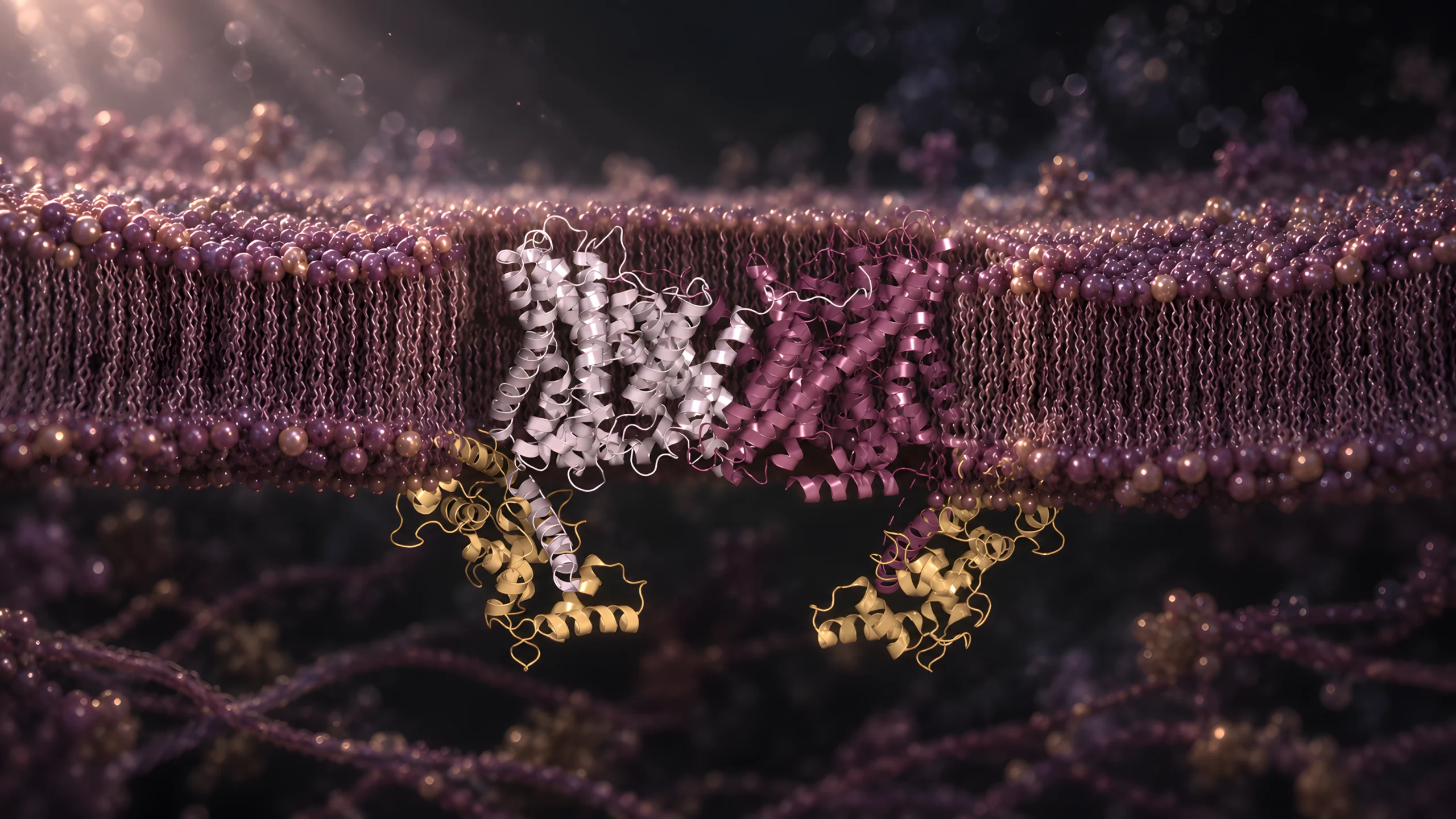
NHE1 is embedded in the plasma membrane enclosing a living cell, with its name referring to sodium-hydrogen exchanger 1. During its usual forward operation, one sodium ion enters the cell as one hydrogen ion leaves. Hydrogen ions are protons; their activity determines pH. Exporting them helps the cell regulate intracellular acidity while contributing to acidification outside.[4]
The exchange is electroneutral. Both ions carry one positive charge, so exchanging them produces no net transfer of electrical charge. A combination of chemical gradients across the membrane supply the driving force, particularly the sodium gradient maintained by the cell’s ion-transport machinery. NHE1 uses that gradient energy without directly consuming ATP with each exchange. This makes it a secondary active transporter. That dependence decides where the exchanger can work. A living keratinocyte keeps its sodium gradient topped up with the ATP-driven sodium pump. A corneocyte, with no metabolism left, cannot. This kind of proton export is work for the living cell, done before it becomes barrier material.
During transport, the protein changes conformation. Human NHE1 assembles as a pair of matching subunits. Each contains a transport core, supported by a scaffold that helps hold the pair together. The core changes position relative to the scaffold, altering access to the region where ions bind.[4]
In the inward-facing form, the binding region is accessible from inside the cell. In the outward-facing form, access is from the outer, extracellular side. This is alternating access: a change in the protein’s conformation exposes the binding region to one side of the membrane, then the other, switching between states as exchange proceeds. Binding and release at opposite sides allow sodium and protons to cross through the exchange cycle. The core’s movement relative to the scaffold gives this mechanism its elevator-like description.[4] It’s extraordinary how machine-like molecular biology can be.
Yanli Dong and colleagues have resolved inward-facing and inhibitor-bound outward-facing forms of the human NHE1 complex.[4] Comparing them shows the transport cores in different positions relative to the scaffold, with the complex embedded in the membrane. Repeated cycles of this molecular movement facilitate proton export from the cell, linking the sodium gradient to changes in acidity on either side. Dong’s group captured the outward-facing form with the inhibitor cariporide bound, and their cryo-EM map located its binding site, showing how specific inhibitors block transport.[4] The mouse experiments later in this article switch the exchanger off with a different inhibitor, HOE694.
A. Inward-facing
7DSV
Extracellular
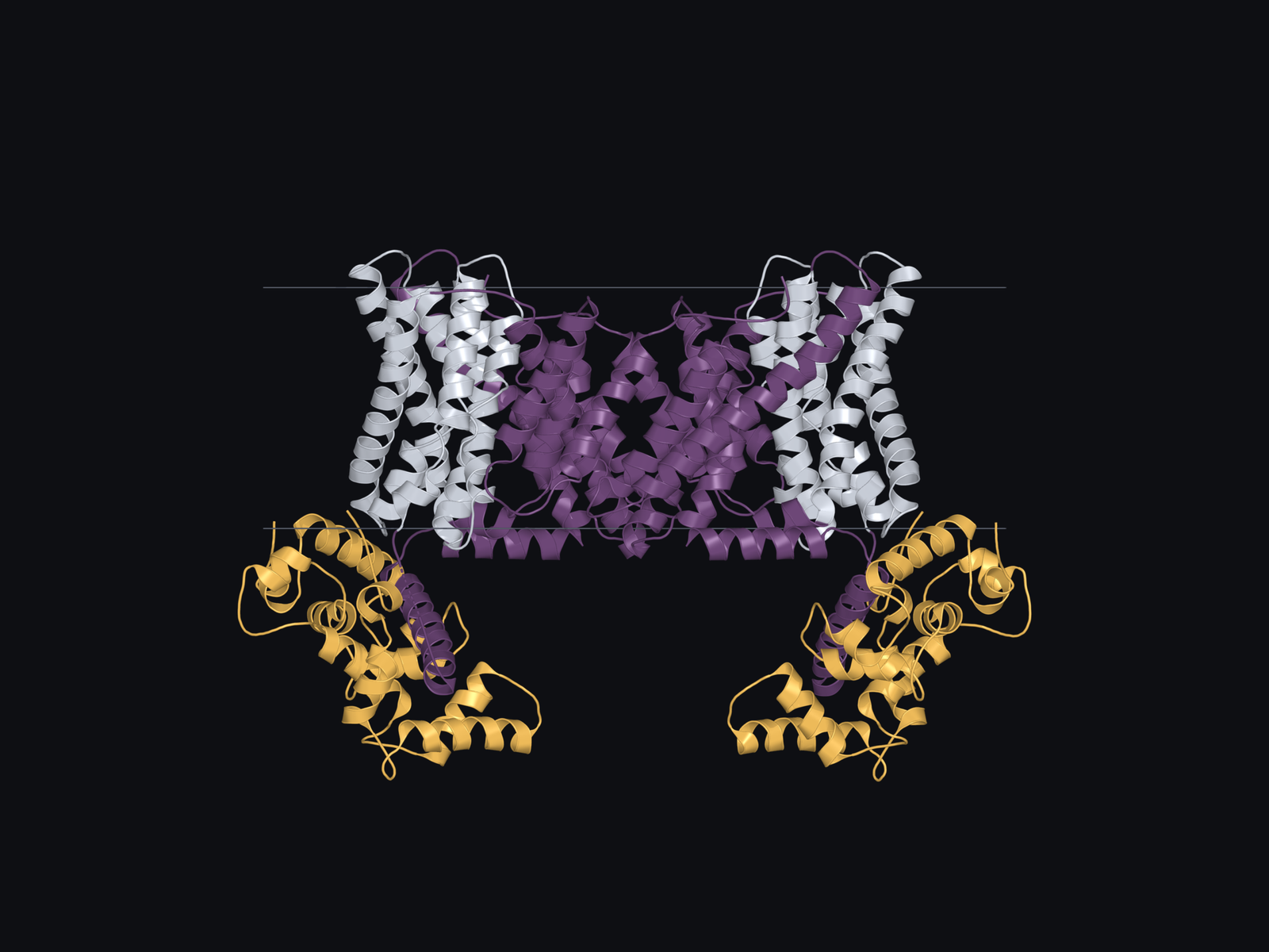
Intracellular
B. Cariporide-bound outward-facing
7DSX
Extracellular
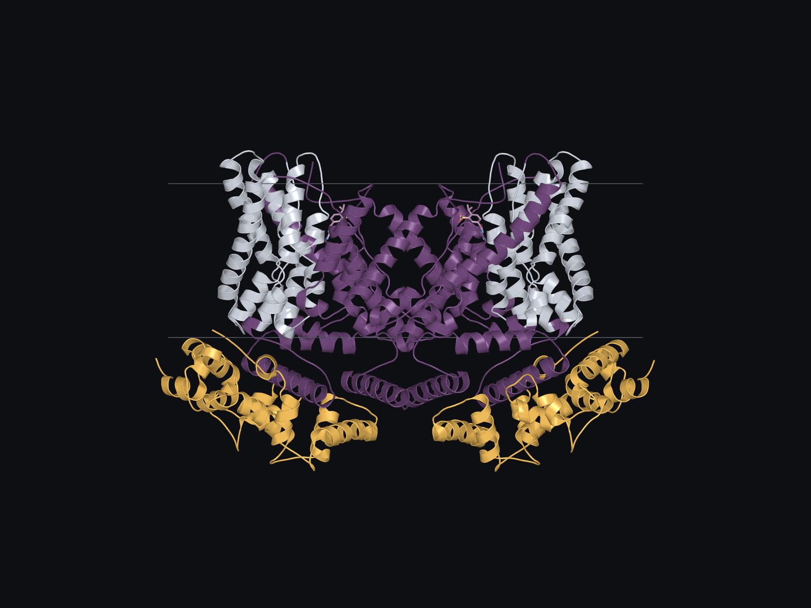
Intracellular
- Transport core
- Scaffold and adjoining NHE1 regions
- CHP1
Human keratinocytes carry this exchanger. Rangaprasad Sarangarajan’s team detected NHE1 in human skin and cultured skin cells, and demonstrated an amiloride-sensitive contribution to intracellular pH regulation in cultured keratinocytes in 2001.[5] NHE1’s presence and contribution to intracellular pH regulation are therefore established in human keratinocytes. Its role in extracellular lipid maturation has been tested most directly in mice.
In mouse skin, NHE1 is found in living granular cells close to the boundary that receives secreted lipids.[6] Extracellular processing begins here, directly outside the cells that carry the exchanger.
The living cell is therefore in a position to influence the surrounding chemical environment beyond its own membrane. As secreted material undergoes conversion, membrane transport helps establish the environment in which that conversion proceeds.
Following acidity into the tissue
Mauro’s experiment, as previously described, had changed the pH from outside the tissue. In 2002, Martin Behne’s group actually removed the exchanger, studying mice lacking NHE1. Conventional measurements at the surface showed only a slight change in pH. Fluorescence lifetime imaging, which maps pH at a much finer scale, looked closer. In the knockout, the acidic extracellular regions at the surface and in the lower stratum corneum had disappeared.[6] The surface reading had concealed the extent of the local change.
What a surface measurement misses. A surface electrode gives a reading at the skin surface. Fluorescence lifetime imaging uses a pH-sensitive fluorescent probe to map acidity within the tissue, resolving local extracellular regions. A modest change in surface pH therefore does not tell us how much acidity has changed around the sites of lipid processing.[6]
These regions were described as acidic microdomains. The term refers to local pockets of acidity within the tissue. A surface electrode cannot resolve these individual pockets, just like a thermometer may give a reading for a room but miss the specific warmth of a radiator. Local acidity can therefore disappear without a large shift in the surface reading.
Mouse epidermis. The study combined mice lacking NHE1 with acute inhibition in barrier-disrupted mouse skin. The acidic rescue tested whether restoring the extracellular environment overcame the inhibitor-associated defects in lipid maturation and recovery.[6]
The researchers also inhibited NHE1 with HOE694 after disrupting the mouse barrier. Lipid secretion continued, while extracellular maturation and barrier recovery were impaired. Applying an acidic buffer overcame the inhibitor-associated defects.[6]
Together, the inhibition and rescue experiments demonstrate that NHE1-dependent acidification supports lipid maturation and barrier recovery in disrupted mouse skin.[6]
NHE1 works in a tissue whose pH varies with location. More than two decades later, in 2024, Keitaro Fukuda’s team used genetically encoded reporters to identify three pH zones in mouse stratum corneum, with a more acidic middle region and less acidic regions above and below it. Reporters displayed outside the cells also detected this zonation.[7]
This newer map complicates a familiar picture of skin becoming steadily more acidic towards its surface. It also leaves the NHE1 intervention experiments addressing a specific question: what happens to local acidity and lipid maturation when this exchanger is removed or inhibited? A complete map of tissue pH requires more than identifying one source of exported protons.
Keeping the exchanger in place
For NHE1 to exchange ions across the cell boundary, the protein has to reach the plasma membrane. Measuring how much protein a tissue contains tells us how much is present. It doesn’t tell us how much is operating there.
Dusko Ilic and colleagues investigated that problem through focal adhesion kinase, or FAK, a protein involved in cellular signalling and organisation. In mice with FAK deleted from keratinocytes, the actin cytoskeleton was disturbed and less NHE1 localised to the plasma membrane. The tissue still contained NHE1, with its abundance initially increased.[8]
Mouse epidermis and cultured mouse and human keratinocytes. The human cell experiments tested NHE1 membrane localisation after FAK depletion. The lipid-processing, barrier-recovery and acidic-rescue findings came from mouse skin.[8]
These mice had impaired acidification, unprocessed extracellular lipids and slower barrier recovery. External acidification rescued recovery. The researchers also linked an intact actin cytoskeleton to NHE1 membrane localisation in their cell experiments. FAK depletion also reduced membrane localisation in cultured human keratinocytes.[8]
FAK participates in many cellular processes, so deleting it doesn’t isolate NHE1 alone. A different component narrows the field: secretory phospholipase A₂, the fatty-acid route into acidity that appears later in this article, was present at similar levels in these mice and their littermates, both at baseline and after barrier disruption.[8] Similar levels don’t guarantee similar activity, as this section has already argued, but they make that route a less likely explanation. With that caveat, the study brings another part of the mechanism into view.
Responding to the conditions outside
The relationship runs back towards the cell. In mice, epidermal NHE1 expression increased within hours of barrier disruption, Jean-Pierre Hachem and colleagues found. They then asked whether the disruption itself, or the accompanying rise in pH, drove the response.[9]
Alkalinising the stratum corneum increased NHE1 expression, while acidification reduced it, independently of barrier status. Acidifying disrupted skin overrode the expression response to the barrier injury. Experiments in cultured keratinocytes also found increased expression in alkaline medium and reduced expression in acidic medium.[9]
Those results show feedback from extracellular pH to transporter expression. Extracellular pH controls how much NHE1 the epidermis makes. In turn, the exchanger helps acidify the space outside the cell, and the acidity of that space adjusts the supply of exchanger. The study measured expression, so it did not establish how the cell detects the change or how much proton transport follows. What it shows is that the supply of NHE1 responds to the environment the exchanger helps influence.
Ageing provides another setting in which this relationship has been examined. Eung-Ho Choi’s group found impaired acidity and barrier homeostasis in moderately aged human and mouse skin. Their mechanistic experiments in mice linked reduced NHE1 expression to defective lipid processing, with acidification correcting the abnormalities.[10] The human findings give the work clinical context, while the causal explanation rests on the mouse experiments.
Part of an overlapping system
NHE1 is one of several routes into epidermal acidity. Processing phospholipids releases free fatty acids, which contribute to both lipid composition and acidification. Joachim Fluhr and colleagues inhibited secretory phospholipase activity in mouse skin, and surface pH rose while the stratum corneum lost integrity and cohesion.[11]
Reconstructed human skin. Filaggrin was reduced in a laboratory-grown human skin construct. The compensatory acidifying responses were followed during development of the construct, alongside measurements of lipid organisation and permeability.[12]
The routes also respond when another contribution is reduced. In Kateřina Vávrová’s reconstructed human skin study, filaggrin deficiency lowered PCA and urocanic acid while pH remained acidic. NHE1 was upregulated first, and by day 14 secretory phospholipase A₂-IIA activity had risen. Ceramide processing was unaffected, but free fatty acids doubled, and the authors linked that excess to less ordered lipid lamellae and higher permeability.[12]
This connects the story to filaggrin without assigning the mechanisms identical jobs. Filaggrin supplies material that is processed into small compounds within corneocytes. NHE1 exchanges ions across living cell membranes. Their contributions reach a shared chemical environment through different reactions and locations.
Acidity also affects more than enzyme activity. Anna Nováčková’s group studied synthetic and extracted human stratum corneum lipids in laboratory systems. Multilamellar assembly required acidic conditions, and neutral conditions changed their organisation and increased water permeability.[13] Those experiments do not establish a direct NHE1 effect in intact skin, but they extend the reason for following pH through the process: the environment influences how lipid molecules organise as well as how their precursors are converted.
At the granular-cornified boundary, membrane transport helps establish acidity around newly secreted lipids. The living cell is preparing the ground for the barrier it will soon become part of.
Lamellar bodies. Membrane-bound organelles in epidermal keratinocytes that carry lipids and enzymes for release into the extracellular space during barrier formation.
Electroneutral. Transport with no net transfer of electrical charge. NHE1 exchanges one positively charged sodium ion for one positively charged hydrogen ion moving in the opposite direction.
Secondary active transporter. A membrane protein that couples movement of one substance to the energetically favourable movement of another. NHE1 uses the sodium gradient to drive hydrogen-ion export, without directly hydrolysing ATP.
Acidic microdomains. Small extracellular regions that are more acidic than their surroundings. The term describes a local chemical environment and does not imply an enclosing membrane.
Actin cytoskeleton. A network of actin filaments that supports cell shape and helps organise movement and positioning of cellular components.
References
1. Holleran, W.M., Takagi, Y., Menon, G.K., et al. (1993). Processing of epidermal glucosylceramides is required for optimal mammalian cutaneous permeability barrier function. Journal of Clinical Investigation, 91(4), pp. 1656-1664. https://doi.org/10.1172/JCI116374
2. Schmuth, M., Man, M.-Q., Weber, F., et al. (2000). Permeability barrier disorder in Niemann-Pick disease: sphingomyelin-ceramide processing required for normal barrier homeostasis. Journal of Investigative Dermatology, 115(3), pp. 459-466. https://doi.org/10.1046/j.1523-1747.2000.00081.x
3. Mauro, T., Holleran, W.M., Grayson, S., et al. (1998). Barrier recovery is impeded at neutral pH, independent of ionic effects: implications for extracellular lipid processing. Archives of Dermatological Research, 290(4), pp. 215-222. https://doi.org/10.1007/s004030050293
4. Dong, Y., Gao, Y., Ilie, A., et al. (2021). Structure and mechanism of the human NHE1-CHP1 complex. Nature Communications, 12, 3474. https://doi.org/10.1038/s41467-021-23496-z
5. Sarangarajan, R., Shumaker, H., Soleimani, M., et al. (2001). Molecular and functional characterization of sodium-hydrogen exchanger in skin as well as cultured keratinocytes and melanocytes. Biochimica et Biophysica Acta, 1511(1), pp. 181-192. https://doi.org/10.1016/S0005-2736(01)00273-5
6. Behne, M.J., Meyer, J.W., Hanson, K.M., et al. (2002). NHE1 regulates the stratum corneum permeability barrier homeostasis: microenvironment acidification assessed with fluorescence lifetime imaging. Journal of Biological Chemistry, 277(49), pp. 47399-47406. https://doi.org/10.1074/jbc.M204759200
7. Fukuda, K., Ito, Y., Furuichi, Y., et al. (2024). Three stepwise pH progressions in stratum corneum for homeostatic maintenance of the skin. Nature Communications, 15, 4062. https://doi.org/10.1038/s41467-024-48226-z
8. Ilic, D., Mao-Qiang, M., Crumrine, D., et al. (2007). Focal adhesion kinase controls pH-dependent epidermal barrier homeostasis by regulating actin-directed Na+/H+ exchanger 1 plasma membrane localization. American Journal of Pathology, 170(6), pp. 2055-2067. https://doi.org/10.2353/ajpath.2007.061277
9. Hachem, J.-P., Behne, M., Aronchik, I., et al. (2005). Extracellular pH controls NHE1 expression in epidermis and keratinocytes: implications for barrier repair. Journal of Investigative Dermatology, 125(4), pp. 790-797. https://doi.org/10.1111/j.0022-202X.2005.23836.x
10. Choi, E.-H., Man, M.-Q., Xu, P., et al. (2007). Stratum corneum acidification is impaired in moderately aged human and murine skin. Journal of Investigative Dermatology, 127(12), pp. 2847-2856. https://doi.org/10.1038/sj.jid.5700913
11. Fluhr, J.W., Kao, J., Jain, M., et al. (2001). Generation of free fatty acids from phospholipids regulates stratum corneum acidification and integrity. Journal of Investigative Dermatology, 117(1), pp. 44-51. https://doi.org/10.1046/j.0022-202x.2001.01399.x
12. Vávrová, K., Henkes, D., Strüver, K., et al. (2014). Filaggrin deficiency leads to impaired lipid profile and altered acidification pathways in a 3D skin construct. Journal of Investigative Dermatology, 134(3), pp. 746-753. https://doi.org/10.1038/jid.2013.402
13. Nováčková, A., Sagrafena, I., Pullmannová, P., et al. (2021). Acidic pH is required for the multilamellar assembly of skin barrier lipids in vitro. Journal of Investigative Dermatology, 141(8), pp. 1915-1921.e4. https://doi.org/10.1016/j.jid.2021.02.014
14. Monteleon, C.L., Agnihotri, T., Dahal, A., et al. (2018). Lysosomes support the degradation, signaling, and mitochondrial metabolism necessary for human epidermal differentiation. Journal of Investigative Dermatology, 138(9), pp. 1945-1954. https://doi.org/10.1016/j.jid.2018.02.035
A short note
The experiments that most directly connect NHE1 to local extracellular acidity and lipid maturation were performed in mice. Human keratinocyte studies establish the transporter’s presence and contribution to cellular pH regulation, while human ageing observations provide a further connection to barrier function. These findings do not assign NHE1 a fixed share of human surface acidity.[5,6,10]
V-ATPase contributes to intracellular acidification and epidermal differentiation, supported by experiments in reconstructed human epidermis.[14] Its contribution to the extracellular environments discussed here remains unresolved. Fukuda’s spatial study also leaves open whether detected V-ATPase components operate in mature stratum corneum.[7]
Behne’s 2002 study reports Sebapharma support for the lead author and an inhibitor supplied by Aventis.[6] The findings concern experimental barrier biology. This article was written independently, without sponsorship, affiliate links or product recommendations.