In skincare, TEWL has become shorthand for almost everything the skin barrier can do wrong. A higher number is read as dehydration, damage and something to correct; a lower one as hydrated, repaired skin. The measurement is useful, but it has been given a to-do list longer than it can fulfil.
Transepidermal water loss, or TEWL, is the rate at which water vapour leaves a defined area of skin, usually reported in grams per square metre per hour. Some outward diffusion is continuous and normal. A healthy barrier regulates that rate, but it doesn’t stop water movement outright. TEWL measures movement. How much water the stratum corneum is actually holding is a separate question. Nor does it inspect the lipids, corneocytes or enzymes controlling that movement. It is a functional readout of water control, not a diagnosis.
The Measurement Happens in the Air
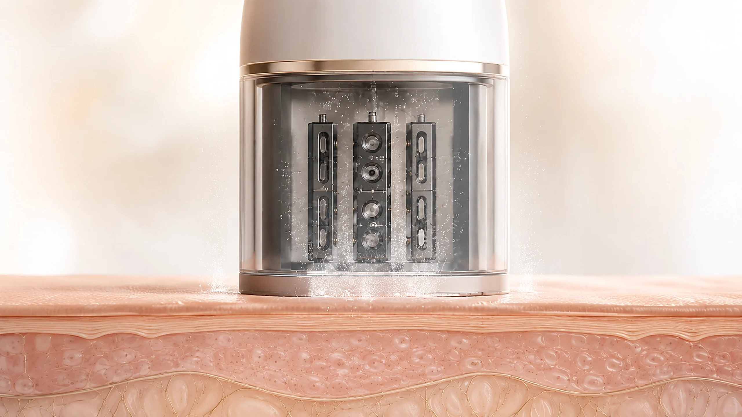
It began in 1977, where a biomedical engineer Gert Nilsson described an instrument for estimating the vapour-pressure gradient immediately above the skin. In this setup a small chamber sits against the skin, open at the top. Water leaves the surface, rises into that chamber, and thins out the further it gets from the skin. Two sensors, positioned at different heights, catch the thinning as a gradient, and the instrument does the arithmetic from there.
Modern TEWL instruments include open, unventilated and condenser-chamber designs, but the original principle survives. The name sounds anatomical, but the measurement happens in the air. A TEWL probe does not count ceramides or inspect lamellar layers. It measures the water that made it out.
Measuring in the air has consequences. Ambient humidity, temperature and airflow can alter the vapour around the probe, while skin temperature, body site and sweating change what leaves the surface in the first place. Researchers therefore allow participants to acclimatise and control the room before treating a small change as biology. Under those controlled, non-sweating conditions, the reading mainly reflects water passing straight out through the stratum corneum. The probe can quantify that flux. It cannot decide why the value changed.
Water Leaving Is Not Water Held
Skin hydration describes how much water is present in the outer skin. TEWL describes how quickly water is crossing it and evaporating. The two can influence each other without ever measuring the same thing.
In 1998, Gil Yosipovitch and colleagues measured 16 healthy volunteers every two hours over a 24-hour period. They tracked TEWL, stratum-corneum hydration, surface pH and skin temperature at four body sites. TEWL followed significant time-dependent rhythms at most sites, with the pattern indicating higher permeability in the evening and night. Stratum-corneum hydration showed no such rhythm, and the two measurements didn’t move together at all. The instruments were asking different biological questions.
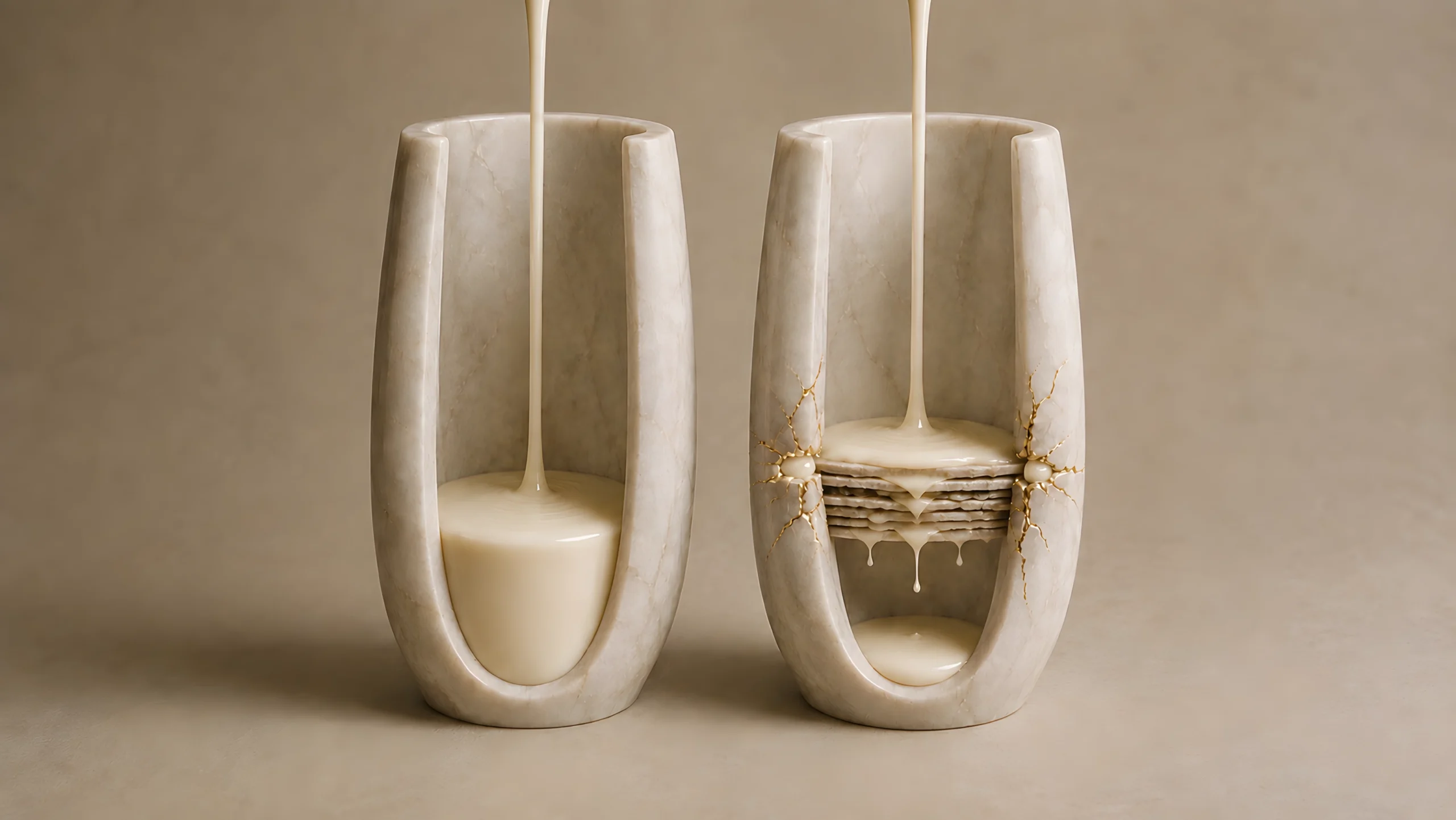
Petrolatum makes the same point from the opposite direction. An occlusive film can lower TEWL quickly by slowing evaporation at the skin-air boundary. The reduced TEWL reading is accurate. What it does not prove is that the skin’s own lipid architecture has already been reconstructed. An external film can change the output before the internal structure changes.
The Number Doesn’t Explain Itself
Under controlled conditions, TEWL is a valuable measure of permeability-barrier function. Joachim Fluhr, Kenneth Feingold and Peter Elias compared readings from multiple devices with absolute water loss determined by weight in human and rodent models. The values tracked one another. When the barrier offers less resistance to water movement, TEWL can detect the change.
The limitation occurs after the reading. An elevated value can follow structural disruption, inflammation, cleansing, heat or a poorly controlled room. The number records that water loss is higher; it does not identify the pressure responsible. A normal value is equally narrow. It cannot prove that lipid organisation, cohesion, inflammatory tone and resilience have all returned. A resting measurement is not a stress test.
A TEWL result becomes useful through making a comparison: the same site before and after a controlled intervention, or carefully matched groups measured under standardised conditions. A single number detached from its site, device and environment has very little to say on its own.
TEWL is valuable because its question is narrow. It tells us how quickly water is escaping from a defined area of skin under defined conditions. It does not measure hydration, locate a damaged structure or name the cause. TEWL is the readout, not the problem. It can show that water control has changed. The biology still has to tell us why.
References
Nilsson, G.E. (1977). Measurement of water exchange through skin. Medical & Biological Engineering & Computing, 15(3), pp. 209-218. https://doi.org/10.1007/BF02441040
Alexander, H., Brown, S., Danby, S. and Flohr, C. (2018). Research techniques made simple: transepidermal water loss measurement as a research tool. Journal of Investigative Dermatology, 138(11), pp. 2295-2300.e1. https://doi.org/10.1016/j.jid.2018.09.001
Yosipovitch, G., Xiong, G.L., Haus, E., et al. (1998). Time-dependent variations of the skin barrier function in humans: transepidermal water loss, stratum corneum hydration, skin surface pH, and skin temperature. Journal of Investigative Dermatology, 110(1), pp. 20-23. https://doi.org/10.1046/j.1523-1747.1998.00069.x
Lodén, M. (1992). The increase in skin hydration after application of emollients with different amounts of lipids. Acta Dermato-Venereologica, 72(5), pp. 327-330. https://doi.org/10.2340/0001555572327330
Fluhr, J.W., Feingold, K.R. and Elias, P.M. (2006). Transepidermal water loss reflects permeability barrier status: validation in human and rodent in vivo and ex vivo models. Experimental Dermatology, 15(7), pp. 483-492. https://doi.org/10.1111/j.1600-0625.2006.00437.x
This article contains no affiliate links, sponsored content or product recommendations. Scientific claims are grounded in the peer-reviewed literature cited above.