You work a cleanser over wet skin, rinse until the lather disappears and reach for a towel. The wash seems finished. Whatever the cleanser lifted from your skin should now be travelling down the drain with it.
Except that rinsing also changes the cleanser. Hard water supplies dissolved calcium and magnesium ions, which associate with certain cleansing molecules and alter how readily they leave the skin. Under controlled washing conditions, that interaction leaves more surfactant behind and weakens the barrier. The evidence for this route into damage is strong; the broader claim that hard water damages skin needs a closer look. [1,5]
Why does water intended to remove a cleanser help it stick? If we can answer that, we begin to understand how the exposure continues after the wash appears to have ended.
Water Changes the Rinse
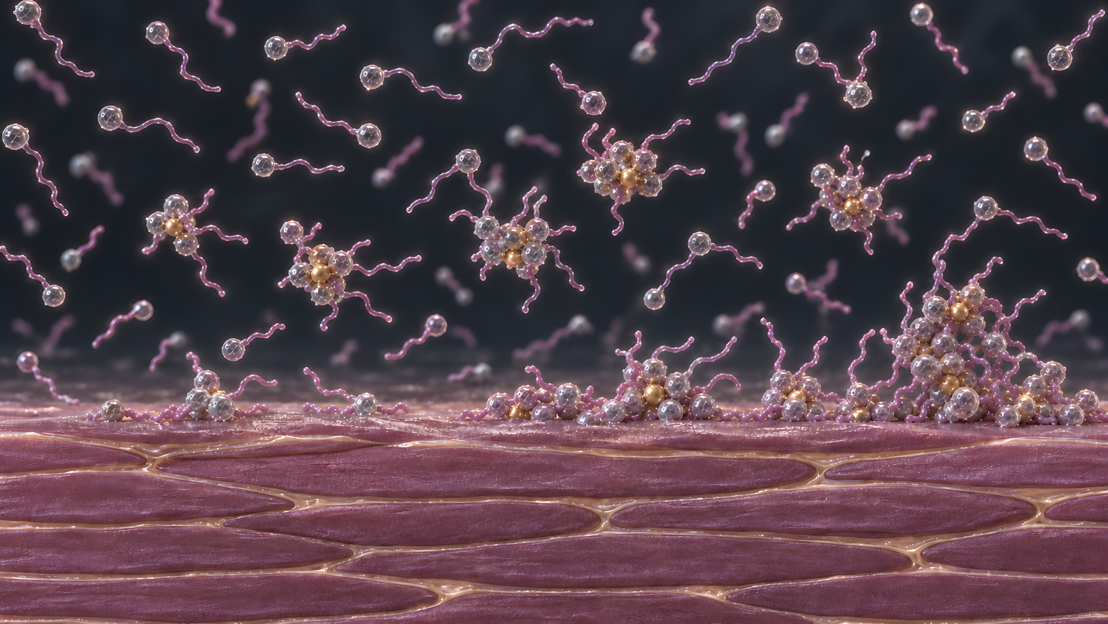
Fatty-acid soap gives us a way into the problem. Its cleansing molecules combine a water-facing, negatively charged head with a tail that associates with oily material. Calcium carries two positive charges. When it binds the fatty-acid anions, it forms calcium soaps that dissolve poorly in water. We recognise this chemistry in soap scum, where cleansing material has combined into something harder to wash away. [1]
That makes a deposit plausible. Soap scum shows the chemistry works in a basin, and a basin isn’t skin. To find out whether the rinse contributes to it, we need to change the rinse water separately from the water used to wash. If calcium in the rinse increases what remains, the removal step has helped create the exposure we are trying to end.
Controlled soap washing on human forearms. Calcium in the wash and rinse waters was varied independently. The experiment identified calcium-soap residue on skin, allowing the researchers to separate the rinse’s contribution from the wash’s. It did not measure subsequent barrier damage.[1]
Nobuki Fujiwara’s team at Mandom made that separation in 1992. They washed human forearms with sodium laurate, a fatty-acid soap, then varied the calcium content of the wash and rinse waters independently. Infrared spectroscopy identified calcium laurate on the skin afterwards. The soap had acquired a different chemical partner, and the researchers could identify the form left behind. [1]
The rinse exerted the greatest influence. The water that was meant to clear the soap away had actually done most of the work of leaving it behind. Calcium-containing rinse water left substantial residue even after washing in calcium-free water, while calcium in the wash followed by a calcium-free rinse left very little. Further rinsing gradually removed the deposit, but calcium in that water slowed its removal. The team proposed that fatty-acid anions already associated with skin reacted with calcium arriving during the rinse. Their measurements supported the explanation without recording every molecular step as it happened. [1]
So a wash in soft water doesn’t necessarily end as a soft-water exposure. The water arriving afterwards still encounters cleansing molecules on the skin, and its calcium changes how they leave. This experiment used concentrated soap and several minutes of immersion, so it describes that particular system. Within it, however, the rinse was chemically active.
Following the Residue into the Barrier
We have found material that survives a rinse. Fujiwara’s measurements stopped there, though. So does it do anything to the skin receiving it?
Simon Danby’s team brought that question into a different cleansing system. Their 2018 study used sodium lauryl sulphate, or SLS, in 80 adults with healthy skin or atopic dermatitis, the common form of eczema. After washing with 10% SLS, very hard water left about 2.8 times as much surfactant on skin as deionised water. Ion-exchange softening reduced the deposit. Adding chlorine did not alter its amount. [5]
Changing the water had changed how much of the same surfactant remained. This extends the retention finding beyond fatty-acid soap, although it does not give SLS the same chemistry as calcium laurate. Danby’s infrared measurements quantified retained SLS without identifying its exact chemical form on skin. The link we can follow is increased retention after rinsing.
A surfactant’s affinity for oily material does not stop at what we want to wash away. The skin’s own lipids surround the flattened, protein-rich cells of the stratum corneum, its outermost layer. Their hydrocarbon chains pack together to help restrict water’s passage. Surfactants interact with these lipids and with skin proteins, so a molecule that remains after rinsing has access to material the barrier needs to preserve. [5,6]
Reading molecular structure. Infrared spectroscopy measures absorption associated with molecular vibrations. Danby’s team used a sulphate signal to quantify retained SLS, and shifts in lipid-chain and protein signals to infer structural changes. Those shifts are chemical measurements rather than images of gaps opening in the barrier.[5]
Danby’s team read changes in that material alongside the residue. After hard-water washing, the lipid signal shifted towards greater disorder, while a protein signal shifted in a direction consistent with denaturation, a change in protein structure. Both shifts correlated with retained SLS. These measurements connect the deposit with changes in the skin’s own organisation, and they appeared directly after the wash. [5]
Controlled SLS challenge in adults with and without eczema. Residue and infrared changes were measured after the 10% SLS wash. Rinsed sites were then covered for 48 hours; irritation and barrier outcomes were assessed 24 hours after the chambers were removed. This separates the immediate measurements from the later response.[5]
The researchers then covered the rinsed sites with chambers for 48 hours, prolonging contact with whatever the wash had left behind. Greater retention tracked with greater redness and higher transepidermal water loss, the outward flow of water through skin. Softening reduced the later impairment. Water crossed the challenged barrier more readily, giving the structural findings a corresponding change in function. [5]
Here the explanation gains strength. Hardness altered retention, retained SLS tracked with disruption of the barrier’s structure, and the prolonged exposure produced greater impairment. Yet the chambers also tell us where to stop. A 10% SLS challenge followed by two days under cover gives the surfactant far longer contact than a wash at home. It demonstrates a route into damage without measuring how much damage a modern cleanser produces during ordinary use.
The receiving skin appeared to change the outcome too. Danby included people with and without common loss-of-function variants in the filaggrin gene. Participants with both eczema and a filaggrin variant showed the largest barrier response, although the study did not detect corresponding differences in the amount deposited between its groups. [5]
Filaggrin helps organise keratin as these outer cells form, then breaks down into water-binding products. Reduced filaggrin function and existing eczema plausibly leave the barrier more susceptible before the wash even begins. The larger response appeared without a detectable difference in residue between groups. Knowing what remained on skin was only part of the explanation; we also needed to know which skin received it.
The Cleanser Changes the Exposure
There’s another reason to pause before carrying the SLS result into your bathroom. A finished cleanser contains a mixture of ingredients, and those ingredients change how its surfactants assemble and encounter calcium.
In water, many surfactants gather into micelles, with their oily tails clustered inside and their water-facing heads exposed. Different surfactants also assemble together. In solution experiments, Kevin Stellner and John Scamehorn found that adding a nonionic surfactant, which carries no charge on its head, increased an anionic system’s tolerance of calcium through mixed-micelle formation. The same mineral encountered a different mixture and produced different behaviour. [2]
Russel Walters’s team at Johnson & Johnson approached the interaction by measuring free calcium. Adding the tested baby cleansers and their surfactant components to water reduced the calcium available as free ions. Calcium remained in the system, but its availability for further interactions had changed. That experiment established a chemical effect; it did not test whether the cleansers protected skin from irritation. [8]
These findings explain why we can’t leave the ingredient mixture out. Calcium’s presence alone can’t predict how much surfactant a particular formula leaves behind. Experiments with skin preparations also show that changing the surfactant mixture changes uptake and barrier effects, although those exposures lasted hours. We need measurements of the finished formulation under the conditions in which people use it to establish the size of its everyday effect. [6,7]
What if we remove the cleanser altogether? A strong direct effect of hardness should also be detectable during water exposure without surfactants, given suitable conditions. Sandrine Bergera Virassamnaik’s team examined that possibility in 2026, repeatedly immersing the forearms of adults with atopic dermatitis but no active flare in soft, hard or chlorinated water. Over three days, the forearms receiving no product routine showed modest, inconsistent changes in water loss, with no consistent separation between water types. [9]
That brief study cannot settle prolonged exposure. It does, however, give little support to a strong effect of hardness alone under the conditions tested. The stronger experimental explanation remains the interaction during washing, where the water helps determine what the cleanser leaves behind.
From a Wash to Eczema
By now, softening the water looks like a plausible way to reduce an exposure. Does that also mean a household softener will improve eczema?
Randomised household treatment trial in children. Both groups continued usual eczema care. Nurses assessing the primary severity outcome were blinded to allocation, although families knew whether they had a softener. The comparison tested the added treatment benefit of softening over 12 weeks.[12]
The proposition needs a test in homes, using eczema itself as the outcome. Kim Thomas’s Softened Water Eczema Trial randomised 336 children with moderate-to-severe eczema to usual care with or without an ion-exchange softener. After 12 weeks, adding softening had produced no benefit in objectively assessed eczema severity or topical medication use. [12]
This result limits the promise we can make. The washing experiments showed that softening reduced surfactant retention and later barrier impairment under a controlled challenge. The household trial asked whether changing the water added enough benefit to improve established disease alongside usual care. For these children, over those 12 weeks, it did not. The mechanism explains an exposure; it does not establish an effective eczema treatment.
Randomised prevention pilot in infants at increased risk. The primary outcome concerned recruitment feasibility. Visible eczema by six months was a secondary clinical outcome, assessed in 67 infants. The pilot was not sized to establish prevention efficacy; its confidence interval included both fewer and more cases with softening.[13]
Prevention asks an earlier question, before eczema develops. Zarif Jabbar-Lopez’s SOFTER pilot installed softeners before birth in households expecting infants with a family history of atopy. Fewer assessed infants developed visible eczema in the softened-water group, but the uncertainty included no benefit and some harm. The pilot established that a larger trial was feasible, leaving prevention unresolved. [13]
For an ordinary wash, the evidence changes what we should pay attention to. The lather disappearing doesn’t tell us how much surfactant has left the skin, and knowing the water is hard doesn’t tell us how a finished cleanser will behave. The formulation and the condition of the receiving barrier help determine the outcome. Hardness is on the list of suspects when we try to understand the wash. On its own, it can’t tell you how your skin will react. The towel comes out, but the wash may not be over.
A Short Note
This article contains no affiliate links, sponsored content or product recommendations. The central evidence concerns the interaction between water and cleansing systems. It does not establish that hard water damages every skin barrier or that a household softener treats eczema.
Water hardness describes mainly dissolved calcium and magnesium, usually expressed as calcium carbonate equivalent. It differs from pH, which describes acidity, and alkalinity, the capacity to neutralise acid. A pH strip cannot tell us how much calcium a cleanser encounters. [3]
Fujiwara’s researchers identified calcium laurate after a sodium-laurate wash. Danby’s team quantified retained SLS and associated infrared changes in lipid and protein structure; they did not directly identify calcium-SLS crystals or map penetration depth. The later irritation test used 48-hour occlusion, with outcomes measured 24 hours after removal. These findings support different parts of the mechanism and should not be collapsed into one continuous observation. The healthy filaggrin-variant group contained only eight participants. Raphael Warren’s 1996 study found drier, redder and less hydrated skin with hard water in its less aggressive washing procedure, with rinse-water hardness exerting the greater influence. Its harsher procedure did not preserve that relationship. [1,4,5]
Several studies had commercial involvement. Fujiwara’s authors worked at Mandom; Warren’s team was affiliated with Procter & Gamble. Harvey Water Softeners funded Danby’s investigator-led work, supplied the test waters and advised on water composition and softening. Walters’s study was funded by Johnson & Johnson, whose Consumer R&D staff authored it. The 2026 water study’s authors were employees of NAOS, the company behind the Bioderma routine it also tested. These interests warrant disclosure alongside the methods and results. They do not, by themselves, invalidate a measurement. [1,4,5,8,9]
Formulation evidence also requires restraint. Less free calcium in a solution does not prove less irritation on skin. Walters’s small human component inferred calcium uptake from its depletion in donor water; it did not demonstrate disruption of the epidermal calcium gradient. James-Smith’s mixture experiments used porcine preparations and exposures lasting hours, while Morris’s human cadaver-skin experiments challenged straightforward accounts of penetration by monomers or intact micelles. These studies support a more detailed view of formulation and exposure, rather than a universal rule about one ingredient. [6,7,8]
The water-only comparison in the 2026 paper was brief and involved small groups; finding no consistent difference does not prove the water types equivalent. Hydration increased and discomfort decreased slightly. Its chlorinated condition exceeded the swimming-pool concentration range quoted by the authors. Product-routine improvements are a separate finding from the untreated-forearm comparison. Hydration and permeability also need separate interpretation: Kaoru Okazaki’s randomised crossover study of 43 nurses found improved hydration and self-assessed hand condition with ultra-pure soft water, without improved measured water loss. [9,10]
Population evidence is mixed. Lopez reported 12% higher adjusted odds of prevalent eczema above 200 mg/L calcium carbonate equivalent, with no association in follow-up incidence. The prospective INMA cohort did not find a childhood eczema association. Henning’s Danish donor study associated harder municipal water with self-reported dryness, including after dermatitis exclusions, but did not measure cleansing residues or barrier permeability. The 2021 systematic review rated the childhood observational association very low certainty; its search ended in June 2020. [11,14,15,16]
SWET received public NIHR funding and equipment and consumables from a water-industry consortium. Its primary assessment was blinded; some small parent-reported benefits came from outcomes where families knew their allocation. SOFTER also received NIHR support, with Harvey supplying and funding the intervention and contributing to design and operations. Investigators retained final design decisions, and the company did not analyse or interpret the results. Visible eczema developed in 12 of 36 assessed infants receiving softened water and 15 of 31 controls. The 15-percentage-point difference had a 95% confidence interval from a 38-point reduction to an 8.3-point increase. [12,13]
Hard water. Water containing relatively high concentrations of dissolved calcium and magnesium ions. Hardness describes these minerals and is separate from water’s pH.
Surfactant. A molecule with regions that interact differently with water and oily material. In cleansers, surfactants help oily material disperse in water so it can be rinsed away.
Ion-exchange softening. A process that removes calcium and magnesium ions from water by exchanging them for other ions. The household softeners discussed here replace them with sodium ions.
Denaturation. A change in a protein’s folded structure. It does not necessarily break the peptide bonds linking its amino acids.
Transepidermal water loss. The passage of water through the epidermis and its evaporation from the surface, usually measured as a rate per unit area. It describes water leaving skin, rather than how much water the skin contains.
Micelles. Assemblies of surfactant molecules in water, with water-avoiding regions gathered towards the interior and water-facing regions exposed outside. Different surfactants can assemble together in mixed micelles.
References
1. Fujiwara, N., Toyooka, I., Ohnishi, K. and Onohara, E. (1992). Adsorption Residue of Fatty Acid Soap on Human Skin: Analysis of Human Skin Surface in situ Using Infrared Spectroscopy. Journal of Society of Cosmetic Chemists of Japan, 26(2), pp. 107–112. https://doi.org/10.5107/sccj.26.107
2. Stellner, K.L. and Scamehorn, J.F. (1989). Hardness tolerance of anionic surfactant solutions. 2. Effect of added nonionic surfactant. Langmuir, 5(1), pp. 77–84. https://doi.org/10.1021/la00085a015
3. US Geological Survey. Hardness of Water; Alkalinity and Water. Accessed 4 October (2026). https://www.usgs.gov/water-science-school/science/hardness-water and https://www.usgs.gov/water-science-school/science/alkalinity-and-water
4. Warren, R., Ertel, K.D., Bartolo, R.G., et al. (1996). The influence of hard water (calcium) and surfactants on irritant contact dermatitis. Contact Dermatitis, 35(6), pp. 337–343. https://doi.org/10.1111/j.1600-0536.1996.tb02414.x
5. Danby, S.G., Brown, K., Wigley, A.M., et al. (2018). The Effect of Water Hardness on Surfactant Deposition after Washing and Subsequent Skin Irritation in Atopic Dermatitis Patients and Healthy Control Subjects. Journal of Investigative Dermatology, 138(1), pp. 68–77. https://doi.org/10.1016/j.jid.2017.08.037
6. James-Smith, M.A., Hellner, B., Annunziato, N. and Mitragotri, S. (2011). Effect of Surfactant Mixtures on Skin Structure and Barrier Properties. Annals of Biomedical Engineering, 39(4), pp. 1215–1223. https://doi.org/10.1007/s10439-010-0190-4
7. Morris, S.A.V., et al. (2019). Mechanisms of anionic surfactant penetration into human skin: Investigating monomer, micelle and submicellar aggregate penetration theories. International Journal of Cosmetic Science, 41(1), pp. 55–66. https://doi.org/10.1111/ics.12511
8. Walters, R.M., Anim-Danso, E., Amato, S.M., et al. (2016). Hard water softening effect of a baby cleanser. Clinical, Cosmetic and Investigational Dermatology, 9, pp. 339–345. https://doi.org/10.2147/CCID.S111729
9. Bergera Virassamnaik, S., Polena, H., Remoué, N., Cadars, B., Prestat-Marquis, E. and Valin, E. (2026). Comparative Effect of Soft, Hard and Chlorinated Water on Atopic Skin and Clinical Benefits of a Dermocosmetic Routine. Cosmetics, 13(3), 159. https://doi.org/10.3390/cosmetics13030159
10. Okazaki, K., Fujino, Y. and Morikawa, Y. (2022). Effects of ultra-pure soft water on the hands of nurses in a neonatal intensive care unit: A randomized crossover study. Contact Dermatitis, 87(6), pp. 521–527. https://doi.org/10.1111/cod.14177
11. Lopez, D.J., Singh, A., Waidyatillake, N.T., et al. (2022). The association between domestic hard water and eczema in adults from the UK Biobank cohort study. British Journal of Dermatology, 187(5), pp. 704–712. https://doi.org/10.1111/bjd.21771
12. Thomas, K.S., Dean, T., O’Leary, C., et al. (2011). A Randomised Controlled Trial of Ion-Exchange Water Softeners for the Treatment of Eczema in Children. PLOS Medicine, 8(2), e1000395. https://doi.org/10.1371/journal.pmed.1000395
13. Jabbar-Lopez, Z.K., Ezzamouri, B., Briley, A., et al. (2022). Randomized controlled pilot trial with ion-exchange water softeners to prevent eczema (SOFTER trial). Clinical & Experimental Allergy, 52(3), pp. 405–415. https://doi.org/10.1111/cea.14071
14. Font-Ribera, L., Gracia-Lavedán, E., Esplugues, A., et al. (2015). Water hardness and eczema at 1 and 4 y of age in the INMA birth cohort. Environmental Research, 142, pp. 579–585. https://doi.org/10.1016/j.envres.2015.07.013
15. Henning, M.A.S., Ibler, K.S., Ullum, H., et al. (2021). The association between water hardness and xerosis: Results from the Danish Blood Donor Study. PLOS ONE, 16(6), e0252462. https://doi.org/10.1371/journal.pone.0252462
16. Jabbar-Lopez, Z.K., Ung, C.Y., Alexander, H., et al. (2021). The effect of water hardness on atopic eczema, skin barrier function: A systematic review, meta-analysis. Clinical & Experimental Allergy, 51(3), pp. 430–451. https://doi.org/10.1111/cea.13797