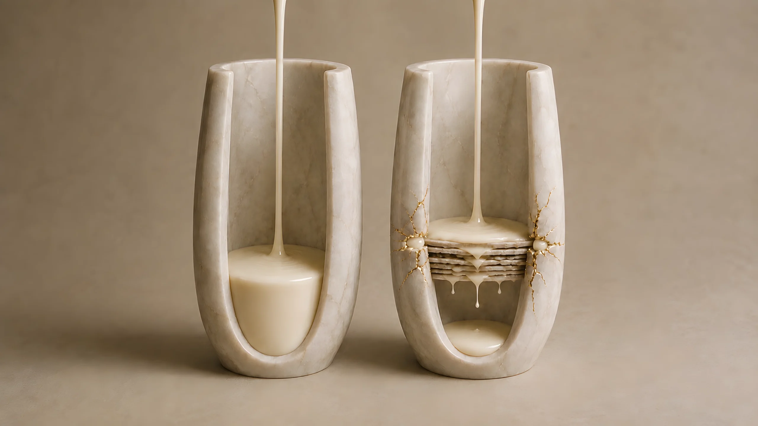
The odd thing about skin-barrier repair is that the cells capable of organising it are located beneath the structure that has failed. The stratum corneum doesn’t have a blood supply, and its corneocytes cannot phone downstairs with a damage report. Yet after an acute disruption, stored barrier material can be released quickly. Lipid metabolism changes over the next several hours. Later, cells in the basal layer increase DNA synthesis and begin expanding the supply of future keratinocytes.
If you’ve ever stood in the mirror wondering whether post-cleanse tightness counts as barrier damage, you will recognise the problem from the other side. You have sensation and a reflection to judge. The living epidermis below has neither. It also doesn’t need them.
An intact stratum corneum maintains a particular environment beneath it. It restricts outward water movement, helps preserve ion distributions and keeps the outer epidermis in a particular physical state. When permeability changes, that state changes too. The skin doesn’t rely on a receptor labelled ‘barrier damage’. It responds to the conditions the failed barrier no longer holds in place.
The Surface Changes the Room Below
IA window doesn’t need to send a memo to your radiator when it opens. The temperature of the room changes, and the heating responds to the temperature it now encounters. Barrier homeostasis works through the same kind of feedback. The event at the surface alters the environment of living cells underneath it.
This sounds obvious after the fact, but it solved a difficult experimental problem. Acetone can remove stratum-corneum lipids. Tape stripping can remove part of the stratum corneum itself. Both also create local injury and can provoke inflammatory responses, so a rise in repair activity after either treatment doesn’t prove that permeability is the regulator. The epidermis might be answering the physical trauma, the chemical treatment or the leak.
The decisive move was to disrupt the barrier and then lend the skin one of its lost functions. If controlling water movement reduced the response while the original structure remained damaged, the feedback could be separated from the injury that created it.
Borrow the Function, Reduce the Response
In 1989, Gabriele Grubauer, Peter Elias and Kenneth Feingold disrupted the permeability barrier of hairless mice and then covered some sites with a water-vapour-impermeable membrane. The covering reduced the expected increase in epidermal lipid synthesis and delayed the native redeposition of barrier lipids. A membrane that allowed water vapour through did not have the same effect. The paper called transepidermal water loss the signal for recovery.
The title captured the experimental logic, though later work would make the word ‘signal’ less exact. The membrane hadn’t rebuilt the stratum corneum. It had acted as a temporary stand-in for one part of its function. While it controlled water loss, the epidermis behaved as though the demand for new lipid had fallen. This is the physical principle behind occlusion, although an experimental latex wrap and a layer of petrolatum aren’t interchangeable treatments.
The connection to why petrolatum works is therefore accurate, but it needs the correct scale. Occlusion can immediately reduce outward water movement. It doesn’t manufacture a new lamellar matrix on contact, and these experiments do not show that ordinary moisturising somehow prevents healthy repair. They show that the repair programme is adjusted to the permeability conditions the epidermis is living under.
Ehrhardt Proksch, Kenneth Feingold, Mao-Qiang Man and Peter Elias then extended the experiment to cell supply. After acetone treatment, epidermal DNA synthesis rose by 102 per cent. Tape stripping raised it by 127 per cent, with the response peaking around 18 to 20 hours and concentrating in the basal layer. A tightly fitted impermeable membrane largely suppressed the increase. Across coverings with different water-vapour permeability, the biological response followed the quality of the borrowed barrier: better water control, less extra DNA synthesis.
Obviously, the tissue wasn’t reading a TEWL meter. TEWL was the researchers’ data from the outside. The living epidermis encountered the accompanying changes in water movement, ions and cellular signalling from within. This became the next question.
Water Loss Was the First Suspect
TEWL rises when the permeability barrier offers less resistance to outward water diffusion. It is a readout of the leak, not a molecule that binds a receptor. Calling it the signal was helpful shorthand for a physical feedback system, but the epidermis still needed a way to translate altered water flux into cellular action.
Seung Hun Lee, Peter Elias, Ehrhardt Proksch and colleagues tested the water-flux idea in 1992. They removed stratum-corneum lipids from hairless mice, then immersed the treated skin in solutions that changed the movement and availability of water and ions. Moving from hypotonic to hypertonic solutions did not support water transit alone as the decisive trigger. Calcium, potassium and phosphate entered the scene.
Calcium or potassium on its own produced a modest inhibition. Together, calcium and potassium reduced barrier recovery from about 50 per cent in controls to between 0 and 11 per cent over two and a half hours. They also prevented the usual increase in HMG-CoA reductase activity and the return of lipid to the stratum corneum. Water movement had not left the story. It had acquired biochemical company.
Calcium Doesn’t Stay Put
In undisturbed epidermis, calcium is relatively low in the deeper basal and spinous layers and rises towards the stratum granulosum, close to the point where living keratinocytes prepare to become corneocytes. The distribution helps coordinate differentiation and lamellar-body secretion. It is closer to a seating plan than a reservoir: where the calcium sits is part of the information.
Gopinathan Menon, Lisa Price, Bommi Bommannan and colleagues altered that seating plan without first removing the barrier. They used high-frequency sonophoresis to manipulate calcium in the upper epidermis of hairless mice while TEWL remained unchanged. When upper-epidermal calcium was displaced back down into the lower layers, lamellar-body secretion accelerated. When calcium was maintained, secretion stayed at its basal rate. The stored material could therefore be released by changing the ionic environment alone. No prior breach was required.
The early model described barrier disruption as a loss of the epidermal calcium gradient. Better imaging later made the account more textured. In 2011, Martin Behne, Susana Sanchez, Nicholas Barry and colleagues used fluorescence-lifetime imaging on unfixed rodent epidermal biopsies. They saw a fast redistribution of calcium after disruption, including mobilisation from intracellular stores and movement between layers and cellular compartments. The pattern changed, but it did not behave as though calcium had drained from one neat extracellular tank.
The refinement is important. Calcium is not a lone alarm wire running from the surface. It is a second messenger distributed across cells, organelles and extracellular spaces. Barrier failure changes the map, and the changed map helps control an early repair event. The Behne study was funded by the European Community’s Marie Curie programme, and the authors declared no conflicts of interest.
Repair Arrives in Overlapping Waves
The first response uses what is already packed. Lamellar bodies in granular keratinocytes contain lipid precursors and processing enzymes. Once secretion is triggered, they fuse with the cell membrane and release their contents at the boundary of the stratum corneum. The material still needs to be processed and arranged into extracellular lamellae, but delivery can begin before the epidermis has made a fresh supply.
The second response changes metabolism. Ehrhardt Proksch, Peter Elias and Kenneth Feingold found that the activation state of HMG-CoA reductase, the rate-limiting enzyme in cholesterol synthesis, changed within 15 minutes of severe acute disruption in mice. Total enzyme activity began rising after around an hour and a half. Walter Holleran and colleagues found a later timetable for sphingolipid synthesis, which peaked five to seven hours after acetone treatment alongside increased serine palmitoyltransferase activity.
These increases were reconstruction rather than general busyness. Kenneth Feingold and colleagues slowed early recovery when they inhibited cholesterol synthesis with topical lovastatin, then restored it with mevalonate or cholesterol. Walter Holleran’s group inhibited sphingolipid synthesis and delayed the later part of recovery; supplying ceramides overcame the delay. The two lipid pathways joined the repair effort on different timeframes.
The third response expands cell supply. The Proksch experiments placed the rise in DNA synthesis later and in the basal layer. Those cells still had to differentiate and move towards the surface before they could contribute mature corneocytes. This is why barrier repair does not share one 28-day clock.
The sequence is economical. Release what is stored, increase the synthesis of what is needed and, if the demand persists, recruit additional cells. The clocks overlap: the first wave holds the breach, the second rebuilds it, and the third stands guard behind it.
What This Means Outside the Laboratory
Most of this framework came from hairless-mouse skin exposed to acetone, tape stripping, sonophoresis or experimental inhibitors. These interventions were designed to make the feedback visible. They do not tell you that one ordinary cleanse produces a 102 per cent rise in DNA synthesis, or that every tight feeling represents a measurable permeability defect. Sensation, TEWL and barrier structure answer related but different questions.
The evidence is also unusually concentrated. Much of the foundational work came from the same University of California, San Francisco and Veterans Affairs research programme across the late 1980s and early 1990s. That continuity allowed one experiment to sharpen the last, but it is not the breadth of independent human replication we would now prefer. No branded barrier-repair product was being compared, and I found no commercial sponsor attached to these foundational experiments. The larger limitation is translation from controlled murine disruption to the smaller, repeated pressures of a human routine.
What the experiments do establish is a feedback sequence. An intact barrier maintains water and ionic conditions. Disruption changes those conditions, calcium is redistributed, preformed lamellar bodies are released, lipid pathways accelerate and the basal layer later increases cell production. The precise mix will change with the insult, but no single switch has sole custody of repair.
So how does skin know its barrier has been damaged? It detects the loss of the state an intact barrier was maintaining.
The surface doesn’t need to send a message down. Its failure changes the message the living epidermis is already receiving.
References
Proksch, E., Feingold, K.R., Man, M.-Q. and Elias, P.M. (1991). Barrier function regulates epidermal DNA synthesis. Journal of Clinical Investigation, 87(5), pp. 1668-1673. https://doi.org/10.1172/JCI115183
Feingold, K.R., Man, M.-Q., Menon, G.K., et al. (1990). Cholesterol synthesis is required for cutaneous barrier function in mice. Journal of Clinical Investigation, 86(5), pp. 1738-1745. https://doi.org/10.1172/JCI114899
Holleran, W.M., Man, M.-Q., Gao, W.N., et al. (1991). Sphingolipids are required for mammalian epidermal barrier function: inhibition of sphingolipid synthesis delays barrier recovery after acute perturbation. Journal of Clinical Investigation, 88(4), pp. 1338-1345. https://doi.org/10.1172/JCI115439
Lee, S.H., Elias, P.M., Proksch, E., et al. (1992). Calcium and potassium are important regulators of barrier homeostasis in murine epidermis. Journal of Clinical Investigation, 89(2), pp. 530-538. https://doi.org/10.1172/JCI115617
Menon, G.K., Price, L.F., Bommannan, B., et al. (1994). Selective obliteration of the epidermal calcium gradient leads to enhanced lamellar body secretion. Journal of Investigative Dermatology, 102(5), pp. 789-795. https://doi.org/10.1111/1523-1747.ep12377921
Behne, M.J., Sanchez, S., Barry, N.P., et al. (2011). Major translocation of calcium upon epidermal barrier insult: imaging and quantification via FLIM/Fourier vector analysis. Archives of Dermatological Research, 303(2), pp. 103-115. https://doi.org/10.1007/s00403-010-1113-9
This article contains no affiliate links, sponsored content or product recommendations. Scientific claims are grounded in the peer-reviewed literature cited above.