CURRENT FOCUS

What if skin type is the wrong question?

Geraint Thomas Bio follows questions like these through the biology underneath them. Right now, the focus is epidermal biology and the science behind skincare, with new topics added as the work develops.

LATEST

Featured writing.

Explore

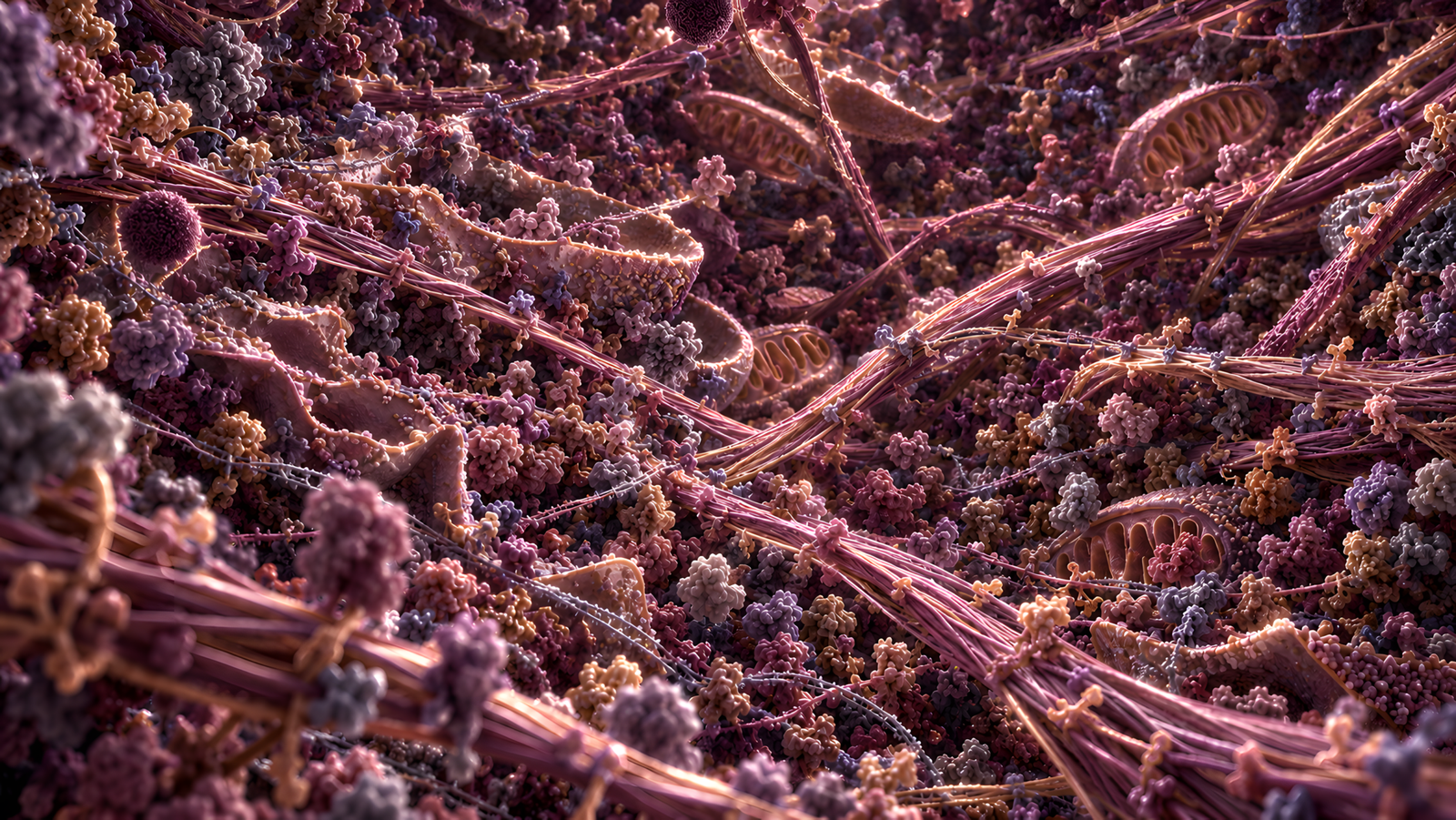
The skin, by scale.

Six stops from the whole organ to a single molecule. Each one has its own biology, and its own ways of going wrong.

≈ 1.8 m²

1 m  ·····  1 m  ·····  1 nm

— 1 m
– 10 cm
– 1 cm
– 1 mm
– 100 µm
– 10 µm
– 1 µm
– 100 nm
– 10 nm
– 1 nm

01 · The organ

The body's boundary.

Roughly 1.5 to 2 m² of surface

Around two square metres of tissue deciding, continuously, what gets in and what stays. Most of that decision is made in the outermost fraction of a millimetre.

≈ 0.1 mm

1 m  ·····  100 µm  ·····  1 nm

– 1 m
– 10 cm
– 1 cm
– 1 mm
— 100 µm
– 10 µm
– 1 µm
– 100 nm
– 10 nm
– 1 nm

02 · The epidermis

Rebuilt from the bottom up.

About 0.1 mm over most of the body

Roughly the thickness of a sheet of paper. New cells form at the base and take around six weeks to reach the surface, changing all the way up.

10–20 µm

1 m  ·····  10 µm  ·····  1 nm

– 1 m
– 10 cm
– 1 cm
– 1 mm
– 100 µm
— 10 µm
– 1 µm
– 100 nm
– 10 nm
– 1 nm

03 · The stratum corneum

Dead, and still working.

15 to 20 cell layers, 10 to 20 µm

The outermost layers are no longer alive in the cellular sense. They still hold water in, keep most things out and shed on a controlled schedule.

< 1 µm thick

1 m  ·····  1 µm  ·····  1 nm

– 1 m
– 10 cm
– 1 cm
– 1 mm
– 100 µm
– 10 µm
— 1 µm
– 100 nm
– 10 nm
– 1 nm

04 · The corneocyte

The brick.

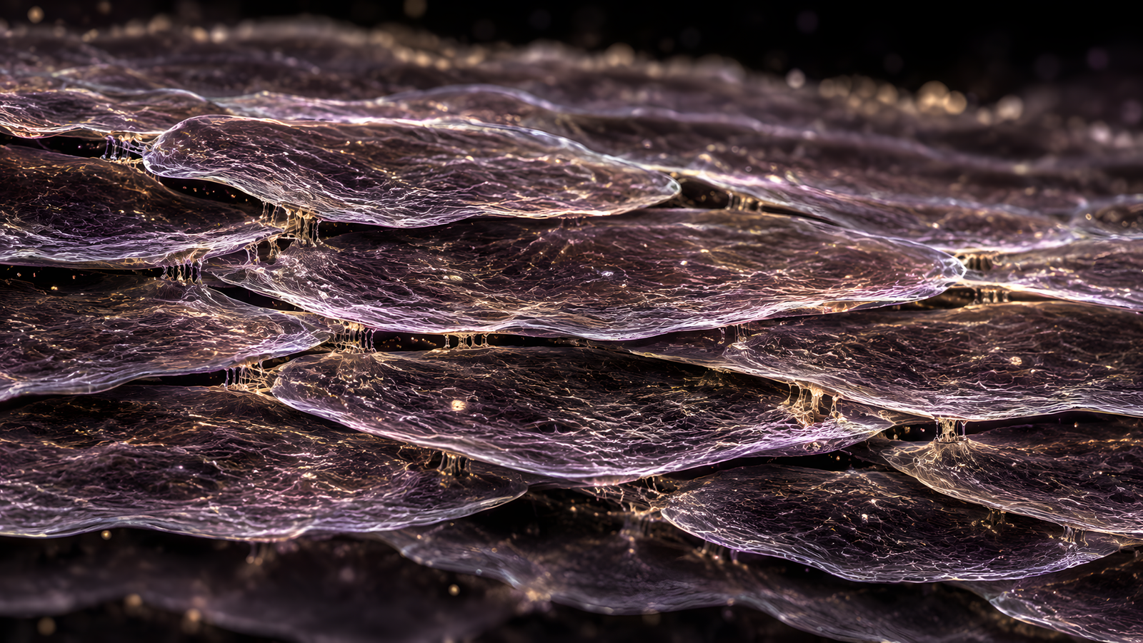
30 to 40 µm across, under 1 µm thick

A flattened cell packed with keratin and sealed inside a cornified envelope. Wide and extraordinarily thin, tiled against its neighbours.

≈ 13 nm repeat

1 m  ·····  10 nm  ·····  1 nm

– 1 m
– 10 cm
– 1 cm
– 1 mm
– 100 µm
– 10 µm
– 1 µm
– 100 nm
— 10 nm
– 1 nm

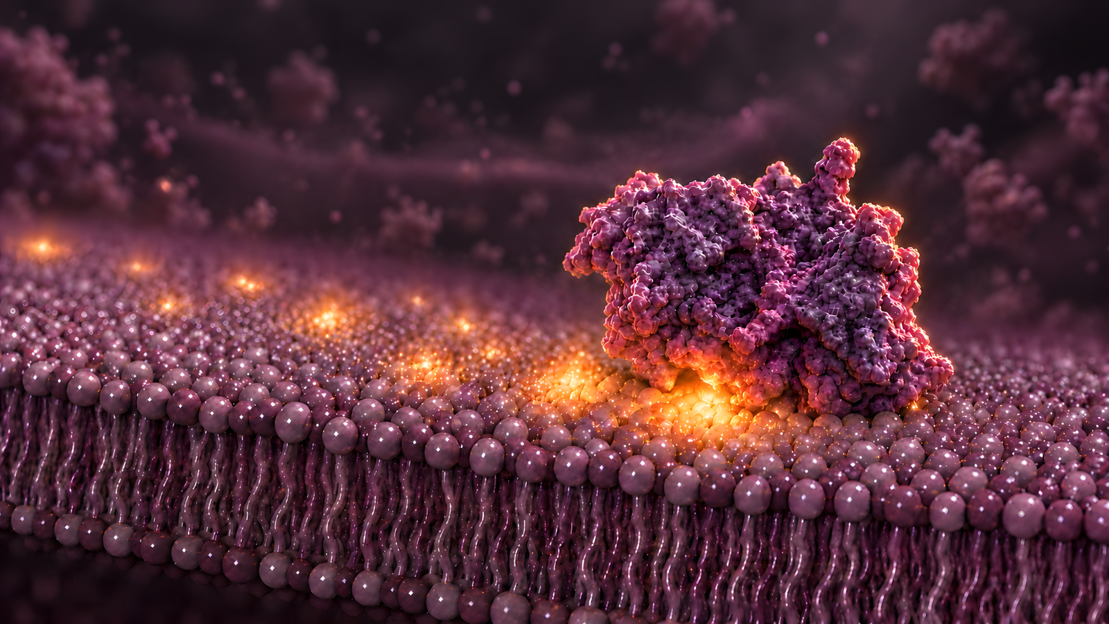
05 · The lipid matrix

The mortar.

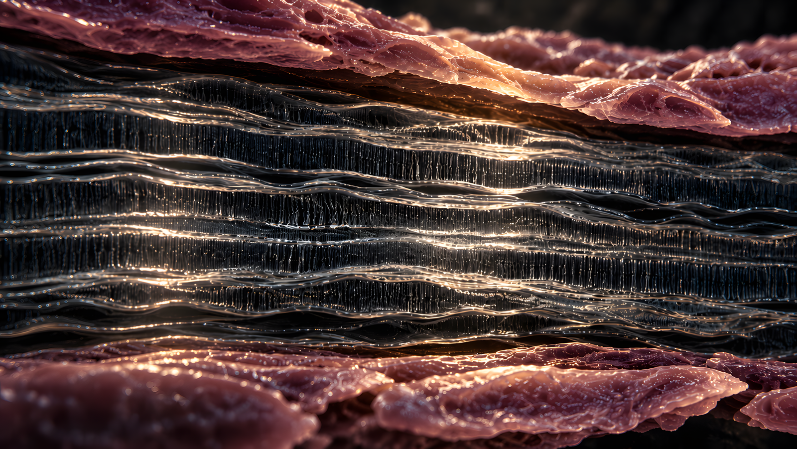
Lamellae repeating at roughly 13 nm

Ceramides, cholesterol and free fatty acids stacked in ordered sheets between the corneocytes. Their organisation sets how quickly water escapes.

≈ 1–10 nm

1 m  ·····  1 nm  ·····  1 nm

– 1 m
– 10 cm
– 1 cm
– 1 mm
– 100 µm
– 10 µm
– 1 µm
– 100 nm
– 10 nm
— 1 nm

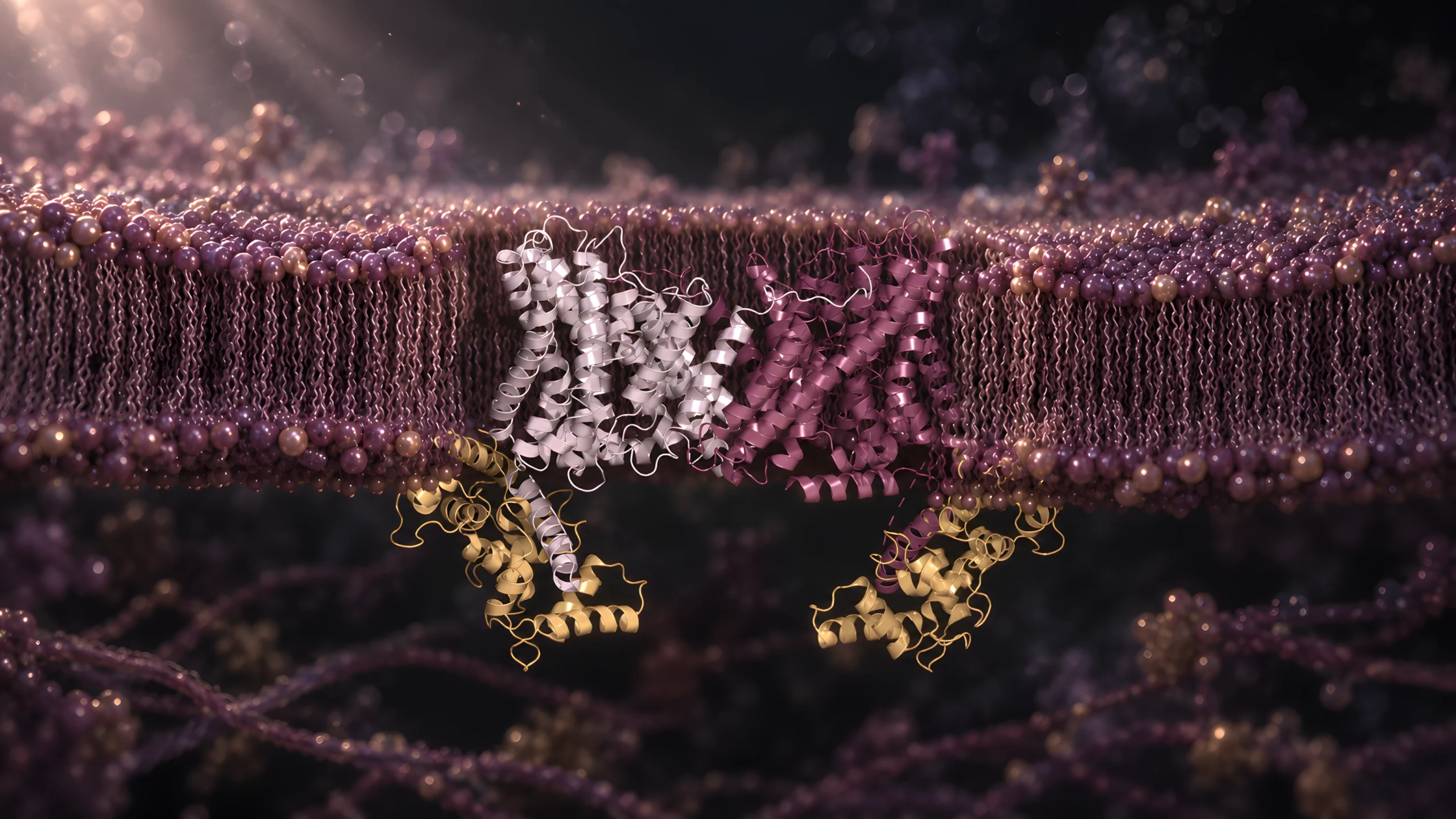
06 · The molecule

Where the barrier is decided.

A few nanometres

Filaggrin, ceramides and the enzymes that process them. Most of what happens at the surface was settled here, long before it arrived.

Scale figures are approximate adult values. Images are illustrations.

EVIDENCE

Trace the claim.

Every claim should have a paper trail.

Find the model. Investigate the method. Evaluate the conditions. Then ask whether the conclusion still holds when it reaches skin.

A result is only as strong as the experiment behind it.

What was actually measured?

In cells, tissue, or people?

Was there a control group?

BOOK

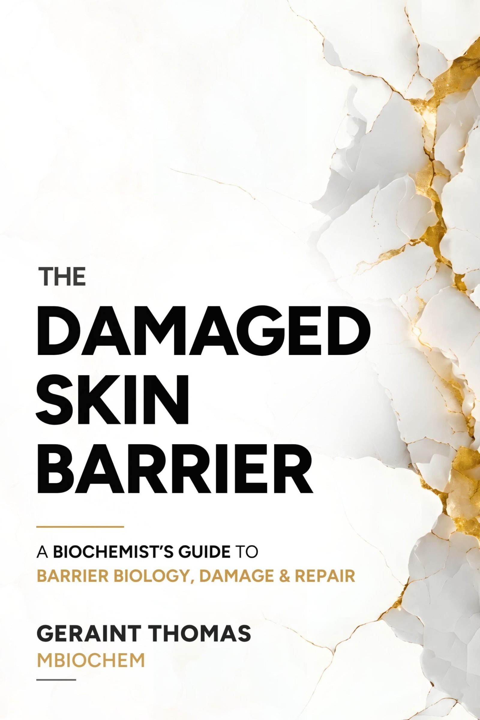
The Damaged Skin Barrier

The long-form version of the questions that started this site. What is the barrier actually made of? How is it regulated? What pushes it out of balance? And what does recovery involve beneath the symptoms we can see and feel?

Written for scientifically curious readers and skin professionals.

252 REFERENCES  ·  24 ORIGINAL  FIGURES  ·  11 CHAPTERS  ·  0 ROUTINES

Cover of The Damaged Skin Barrier: A Biochemist's Guide to Barrier Biology, Damage and Repair, by Geraint Thomas

BACKGROUND

Biochemistry before biography.

I’m a biochemist interested in how biological systems behave, adapt, and more so, when and why they fail.

My research background is in cancer-cell biology and apoptotis. More recently, that same way of thinking has led me into epidermal biology and the science behind skincare.

EXPLORE

Follow the mechanism.

Start with a question, move into the biology, or go straight to the sources. The site is organised so each route connects back to the evidence.