CURRENT FOCUS
What if skin type is the wrong question?
Geraint Thomas Bio follows questions like these through the biology underneath them. Right now, the focus is epidermal biology and the science behind skincare, with new topics added as the work develops.
evidence in context
barrier signalling
measurement
Latest
Ways into the science
01 / Foundations
Scratch the surface.
Start with the core biology behind the subject.
02 / Mechanisms
Under the skin.
See how cells, signals, lipids and environmental inputs interact.
03 / Deep dives
Down the rabbit hole.
Take one question through the evidence and the competing explanations.
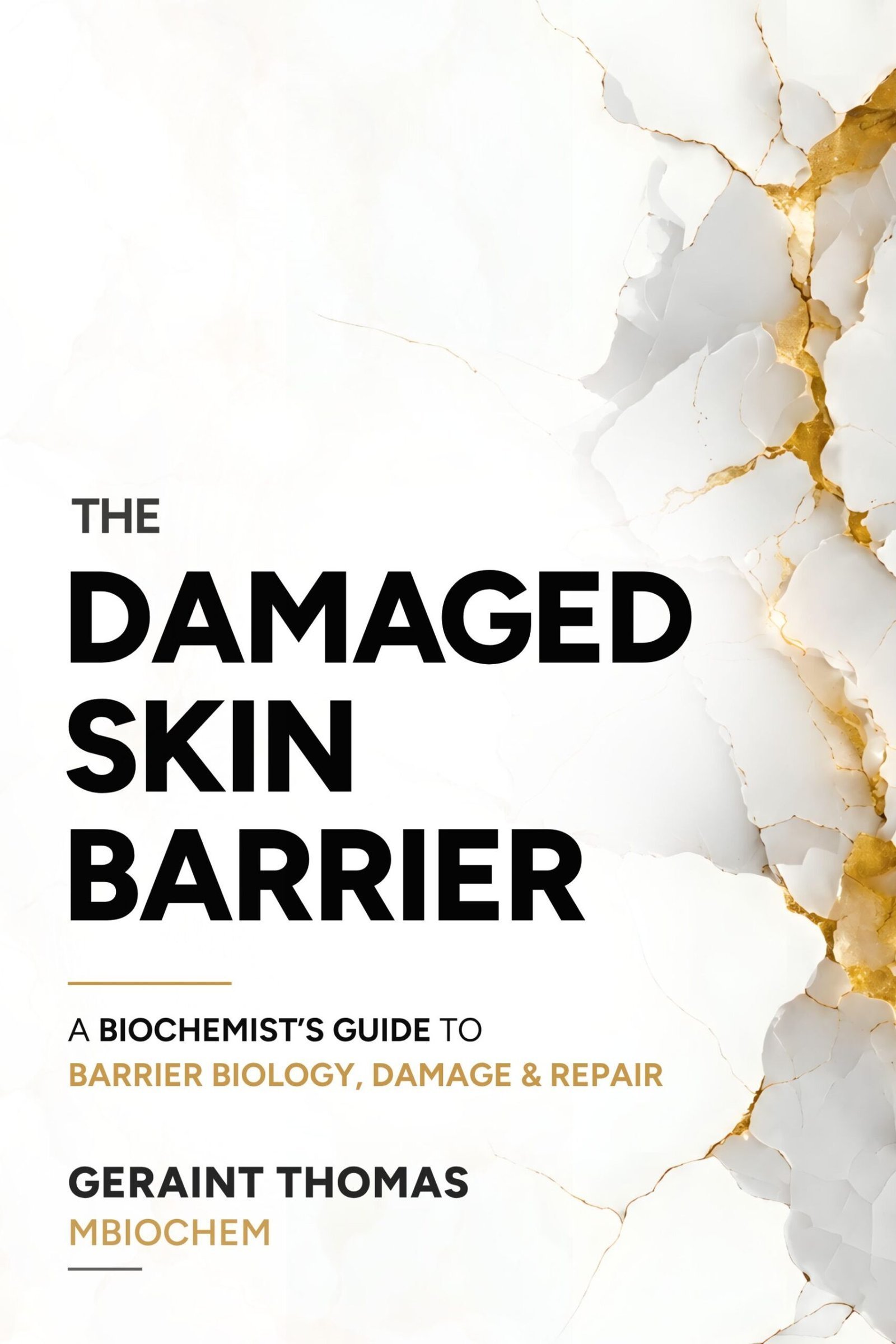
The Damaged Skin Barrier
The long-form version of the questions that started this site.
Written for scientifically curious readers and skin professionals.
01 / STRUCTURE
What is it made of?
Corneocytes, organised lipids and the adhesive structures that hold the sheet together.
02 / REGULATION
How is it regulated?
Surface pH, enzyme activity and a calcium gradient coordinate renewal.
03 / PRESSURE
What pushes it out of balance?
Cleansing, actives, friction, UV and hard water, accumulating on one tissue.
04 / RECOVERY
What does recovery involve?
Feeling better can arrive before the structure is rebuilt.
The skin, by scale.
Six depths, from the whole organ to a single molecule. Select one to zoom in.
Zoomed in · illustrative, not to scale
01 · The organ
The body's boundary.
Roughly 1.5 to 2 m² of surface
Around two square metres of tissue deciding, continuously, what gets in and what stays. Most of that decision is made in the outermost fraction of a millimetre.
02 · The epidermis
Rebuilt from the bottom up.
About 0.1 mm over most of the body
Roughly the thickness of a sheet of paper. New cells form at the base and take around six weeks to reach the surface, changing all the way up.
03 · The stratum corneum
Dead, and still working.
15 to 20 cell layers, 10 to 20 µm
The outermost layers are no longer alive in the cellular sense. They still hold water in, keep most things out and shed on a controlled schedule.
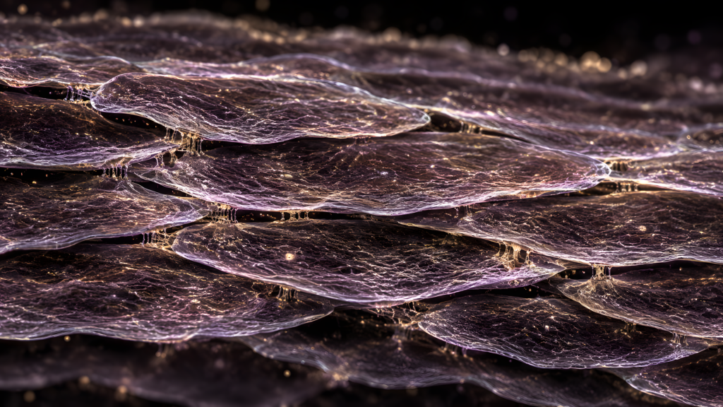
04 · The corneocyte
The brick.
30 to 40 µm across, under 1 µm thick
A flattened cell packed with keratin and sealed inside a cornified envelope. Wide and extraordinarily thin, tiled against its neighbours.
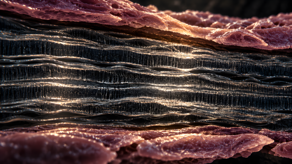
05 · The lipid matrix
The mortar.
Lamellae repeating at roughly 13 nm
Ceramides, cholesterol and free fatty acids stacked in ordered sheets between the corneocytes. Their organisation sets how quickly water escapes.
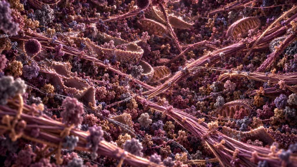
06 · The molecule
Where the barrier is decided.
A few nanometres
Filaggrin, ceramides and the enzymes that process them. Most of what happens at the surface was settled here, long before it arrived.
Scale figures are approximate adult values. Images are illustrations.
Every claim should have a paper trail.
A result is only as strong as the experiment behind it.
Choose a claim, then select an underlined phrase. Each question shows what would strengthen the claim and what would still leave doubt.
.
Select an underlined phrase to test what the evidence supports.Was the difference inherited?
A genotype measured independently of active skin disease, with the variant’s frequency compared in people with and without eczema.
Reduced filaggrin staining in an inflamed lesion. Inflammation can itself lower filaggrin expression.
Which variants?
Clearly defined loss-of-function variants in the FLG gene, with the variants tested stated for the population studied.
A vague label such as “low filaggrin” that mixes gene variants, protein abundance and assay results.
How much risk?
An effect estimate with uncertainty, preferably replicated in another cohort. Genetic association supports a role in susceptibility.
Treating a higher risk as proof that the variant alone causes eczema in every carrier.
Which disease and population?
A stated diagnostic definition and ancestry or population context; variant frequencies and effects differ across populations.
Applying findings from one set of variants or one cohort to all people with atopic dermatitis.
.
Select an underlined phrase to test what the evidence supports.Did the compound act through protein X?
Show target engagement, then corroborate the result with an independent inhibitor or genetic perturbation; a rescue experiment is stronger still.
Assume a compound is selective because its label names protein X. Off-target effects can produce the same result.
What does “sensitises” mean here?
Compare TRAIL alone, inhibitor alone and the combination across doses. Claim synergy only after testing the combination against a defined additivity model.
Call the combination synergistic simply because it has a larger effect than either treatment alone.
Which cells were tested?
Identify the cell models and repeat the finding in independent models, including relevant non-malignant cells where selectivity is part of the claim.
Generalise from one immortalised cell line to “cancer cells” as a whole.
How was the response measured?
A defined viability assay, suitable controls and independent repeats can show whether the inhibitor changes the cells’ response to TRAIL.
An unreplicated result, or an assay without clear controls and normalisation, makes the size of the effect hard to judge.
.
Select an underlined phrase to test what the evidence supports.How was “high” defined?
Use a prespecified threshold, or develop it in one dataset and evaluate it in a separate cohort. Analyse the continuous measurement too when possible.
Choose the cut-off that gives the smallest P value in the same patients and present it as established.
What was measured, and in which cells?
Describe the assay, its reproducibility and whether the signal comes from tumour cells or surrounding tissue.
Treat bulk RNA from mixed tissue as a direct measure of protein abundance in tumour cells.
Association or useful prediction?
Test whether the marker adds information beyond established factors such as stage, then validate the model in new patients.
Use a single unadjusted association to imply the marker can predict an individual patient’s future.
What outcome, and with what precision?
Define overall survival and follow-up; report an effect estimate with a confidence interval and numbers at risk.
Say “worse prognosis” without a defined endpoint, adequate follow-up or an estimate of uncertainty.
How many independent observations?
Three wells from one cell preparation do not make three independent experiments. Count the independent cultures, samples or people.
Could expectation affect the result?
Blinding matters when a person scores an image, stain or lesion. Ask whether the scoring rule was set in advance.
Has the result been replicated?
Independent replication in a new cohort, cell model or lab helps show whether the finding is reliable beyond the original experiment.
Evidence behind these examples: FLG genetics · inflammation and filaggrin · drug combinations · cell-death assays · REMARK guidance. The drug and biomarker claims are illustrative, not reports of a particular study.
Hello, I'm Geraint.
I’m a biochemist interested in how biological systems behave, adapt, and more so, when and why they fail.
I took an integrated master’s in biochemistry at Cardiff University and graduated with a First. My research year was at the European Cancer Stem Cell Research Institute, studying how breast cancer stem-like cells resist death-receptor signalling.
After that I spent several years outside science. Skin biology is what brought me back.
The way of thinking carried over: what was measured, in what, and what does the mechanism say? The epidermis turned out to be a good place to use it. It became The Damaged Skin Barrier and this site. I wrote the book, drew its figures and built the platform myself.

Where next?
Read, check a source, or meet the person behind it. Every route leads back to the evidence.